LAB 7.1 Heart & blood vessels
PLAY VIDEO
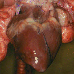 A pig's heart
-
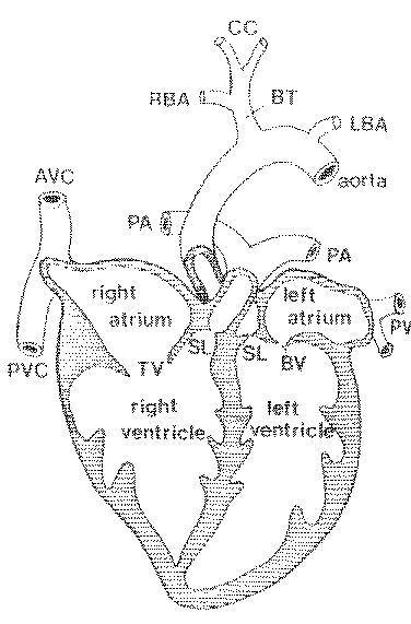
The right ventricle pumps blood through the
semilunar valves and into the pulmonary arteries and then to the lungs.
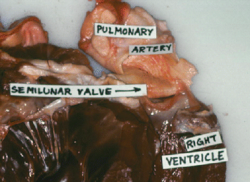 Here are the bovine
semilunar valves.
Here are the bovine
semilunar valves.
-
Oxygenated blood returns to the left atrium
in the pulmonary veins, through the bicuspid valve.
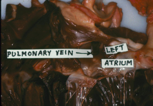 Here is the bovine pulmonary
vein.
Here is the bovine pulmonary
vein.
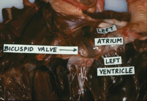 Here is the bovine bicuspid
valve.
Here is the bovine bicuspid
valve.
-
The atrium fills the left ventricle, and
oxygenated blood is then pumped through the aorta to the body
tissues.
 Here is the bovine aorta.
-
The aorta branches to form the major arteries. These
branch again many times and eventually give rise to arterioles and,
finally, to capillaries.
-
Blood is collected from the body tissues by the venous
system,
and eventually returns to the right atrium via the anterior vena
cava and the posterior vena cava for another cycle through the
lungs. Thus,
relative to other arteries, the pulmonary artery is unusual because it
contains de-oxygenated blood. And, relative to other veins, the
pulmonary vein is unusual because it contains oxygenated blood.
-
In the arterial system of meat animals, the aorta bends to
the LEFT
side of the body,
and then runs posteriorly in the midline, ventral to the vertebral
column. It supplies arterial blood to all the body except the lungs.
-
In the arterial system of poultry, the aorta swings to the
RIGHT
side of the body after leaving the heart .
-
If poultry develop heavy muscling, the thin-walled
right ventricle may be unable to cope with the increased cardiovascular
demands and the ventricular wall may thicken as it adapts to the
situation. But this may prevent the atrioventricular valve from
functioning properly and the back pressure to the liver may cause
ascites, an accumulation of fluid in the abdomen that may kill the bird.
-
The venous system of meat animals is dominated by the
anterior
and posterior vena cava.
-
The venous system of poultry is distinguished by a loop in
the neck created by the jugular anastomosis and a loop around the
kidneys.
Cardiac muscle fibres. There
are three different types of muscle tissue in the body
-smooth, cardiac and skeletal.
Smooth muscle occurs in the digestive and reproductive tracts, cardiac
muscle is only found in the heart, while skeletal muscle forms all the
meat of the commercial carcass. Most cardiac muscle cells are
mononucleate. They are arranged in
rows
to form branching fibers, but individual cells are separated by
intercalated discs. Cardiac muscles have a striated appearance due to
the precise alignment of sliding filaments in their contractile
fibrils, but skeletal muscles also are striated. Transverse striations are detailed in
LEC 11.
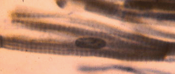 This is a
lateral view of a squashed cardiac muscle cell. The nucleus is fairly
obvious, as are the transverse striations, but it is difficult to see
how this was once part of a branched cardiac muscle fiber.
This is a
lateral view of a squashed cardiac muscle cell. The nucleus is fairly
obvious, as are the transverse striations, but it is difficult to see
how this was once part of a branched cardiac muscle fiber.
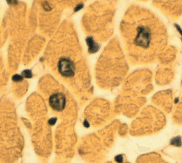 This is a transverse section of a group of cardiac muscle
fibers, showing how their nuclei are located centrally in the axis of
each fiber.
This is a transverse section of a group of cardiac muscle
fibers, showing how their nuclei are located centrally in the axis of
each fiber.
Action potentials.
Cardiac muscle cells are continuously pumping out sodium ions
through their membranes. This causes the inside of the cell to have an
electrical charge of approximately -90 mV with respect to the outside
of the cell. This is called a resting potential. Extrinsic factors such
as electrical activity (ionic movements) in adjacent cells may decrease
the resting potential towards zero. When it reaches a value of
approximately -65 mV, the threshold potential, the decrease in
electrical potential accelerates, and it shoots past the zero value so
that for a brief instant (about one tenth of a second) the membrane
potential is positive. This sudden reversal of electrical charges is
called an action potential. Action potentials are propogated into the
interior of cardiac muscle cells by transverse tubules. In each cell,
the transverse tubular system is an extensive series of finger-like
indentations of the surface membrane.
Cardiac contraction.
This is the same as the
contraction of skeletal muscle - which is described in LEC 11.
The arrival of an action potential in the interior of the
cardiac
muscle cell causes the release of calcium ions from the sarcoplasmic
reticulum. The sarcoplasmic reticulum is a series of membrane-bounded
vesicles in the interior of the cell. Unlike the transverse tubular
system, the sarcoplasmic reticulum does not open to the surface of the
muscle cell. Units of the sarcoplasmic reticulum surround the
contractile fibrils in the interior of cardiac muscle cells. The
sarcoplasmic reticulum sequesters and stores calcium ions, but it
releases them again when prompted to do so by the transverse tubular
system. Calcium ions activate the system of sliding protein filaments
which is responsible for muscle contraction.
Intrinsic rhythm. The
intrinsic rhythm of heart contraction originates from a group of
cells at the sinu-atrial node. The membranes of these cells behave as
if they had a sodium ion leak. Thus, at regular intervals their resting
potentials drop to their threshold values, and they initiate action
potentials. Action potentials then spread in a coordinated wave through
right and left atria. The atria then contract and pump blood into the
ventricles. However, the ventricles also are capable of filling
themselves as they expand after pumping out their previous fill of
blood. Under normal conditions, atrial contraction contributes to the
overall cardiovascular efficiency, but its contribution may become
vital when the heart is weakened by disease. In the medial wall of the
heart, at the junction between the atria and ventricles, is a sensitive
group of cells forming the atrioventricular node. This node is
connected to a conduction system called the bundle of His that runs
down the medial wall separating left and right ventricles. The
atrioventricular node is activated by contraction of the atrial cells,
and the bundle of His conducts the action potential wave to the base of
the ventricles. From this point, a wave of contraction spreads upwards
through the ventricles so that the the blood that has just filled the
ventricles is now pumped out of the heart. The intrinsic heart rate is
determined by the rate at which the sinu-atrial cells "leak" or
depolarize, by the value of their threshold potentials, and by their
resting potential. The flow of blood through the heart is directed by
the heart valves. Mitral
and tricuspid valves make a "lub" sound and the semilunar valves make a
"dup" sound.
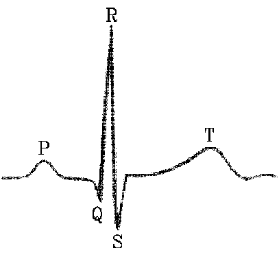
Electrocardiogram (ECG).
Coordinated electrical activity of cardiac muscle cells generates an
electrical signal that may be detected on the surface of the fore flank
as an electrocardiogram.
The P wave is due to atrial excitation,
PQ is the delay as the action potential passes down the bundle
of His,
QRS is due to ventricular contraction or systole, and
T is caused by repolarization of the ventricles.
Control of heart rate. Activity
of the heart is greatly influenced by
its ionic environment. Isotonic sodium chloride plus calcium ions tend
to stop the heart in systole (contracted) while isotonic sodium
chloride plus potassium ions tend to stop the heart in diastole
(relaxed).
The nervous system also has an effect on heart rate. The
thoracic nerve of the sympathetic nervous system releases
catecholamines that increase the heart rate (tachycardia) while the
vagus nerve of the parasympathetic nervous system releases
acetylcholine that slows the heart (bradycardia). The neural regulation
of cardiac activity is a reflex response to inputs from blood pressure
receptors or baroreceptors, and from chemoreceptors that monitor the
concentration of carbon dioxide in the blood. When the heart contracts,
it works against the resistance to blood flow created by the peripheral
blood vessels in the body tissues. Thus, if the peripheral blood
vessels decrease their diameter (vasoconstriction), the blood pressure
tends to rise. Conversely, if peripheral blood vessels are dilated
(vasodilation), the blood pressure tends to drop.
Study hint: university exams
always contain some keener's questions to reward students who are
REALLY keen. In this course, the real keeners are usually vet
wannabees and they are usually really keen to learn the names of the
major blood vessels given on the links below.
The same information is on the web
(http://www.aps.uoguelph.ca/~swatland/gasman.htm) and in a text book
(Structure and Development of Meat Animals and Poultry).
Major arteries of meat animals.
Major arteries of poultry.
Major veins of meat animals.
Major veins of poultry.