-
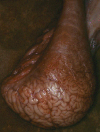
Female mammals have a pair of ovaries located posteriorly and dorsally
in the abdominal cavity Figure 1-21c.The image
below shows one ovary of a cow.
-
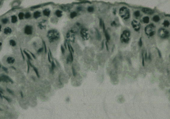
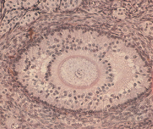
Ova develop in the cortex (outer layer) of the ovary. Each ripe
ovum is enclosed in a fluid-filled follicle, as shown in the histological
image below.
At estrus, ova are released into a ciliated funnel or infundibulum
at the end of each oviduct (fallopian tube).
-
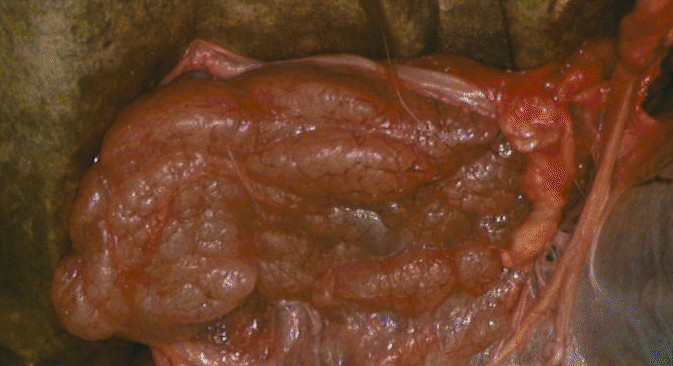
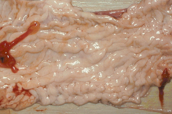
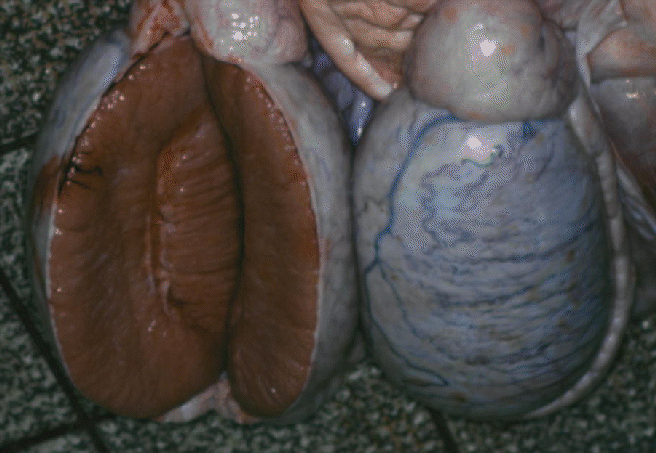
The oviduct on each side leads into a horn of the uterus where embryonic
development takes place. The image below shows one horn of the uterus of
a cow.

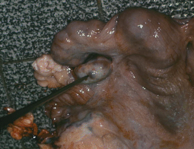
At birth, the offspring emerge through the dilated cervix and
vagina.The
image below shows an undilated cervix of a cow.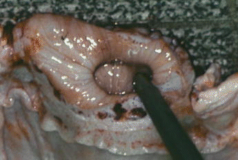
-
Mammary
glands are derived from highly modified sweat glands of the skin.
The udders of sheep and goats are divided into right and left halves,
each with a teat. The cow's udder has four quarters so that there are two
teats on each side. Most sows have seven pairs of mammary glands and a
total of 14 teats. Milk is produced in glandular alveoli, and it
collects in the cistern of the teat. The bovine udder is supported
by medial and lateral suspensory ligaments which are dominated by elastin
and collagen fibers, respectively.
-
In female poultry Figure 1-21d. there is only
a single ovary since the ovary and oviduct of the right side
do not normally develop.
-
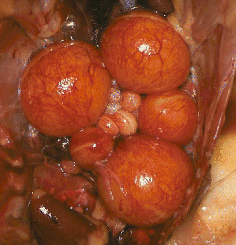
In poultry, the ovary usually contains a cluster of ova in different stages
of development, as shown below.
-
The ova in the most advanced state of development appear as full-sized
egg yolks. A large infundibulum (ostium) leads to a thick
glandular
region of the oviduct where egg albumen is formed, then to a narrower isthmuswhere
shell membranes are added, and finally to a wide uterus where a
calcareous shell is formed.Here is the glandular region, with the oviduct
slit open and laid out flat.
 The vagina opens into
the cloaca and forms mucus to facilitate egg-laying.
The vagina opens into
the cloaca and forms mucus to facilitate egg-laying.
 Here are two boar testes, the one on
the left sliced open.
Here are two boar testes, the one on
the left sliced open.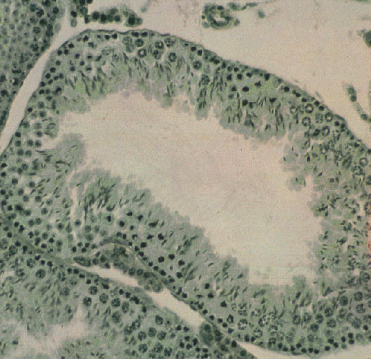 The image
above shows one tubule under the microscope. The higher power image below
shows part of the tubule (lumen downwards) with meiotic divisions
leading to the formation of spermatozoa, whose tails can be seen faintly
at the bottom of the image.
The image
above shows one tubule under the microscope. The higher power image below
shows part of the tubule (lumen downwards) with meiotic divisions
leading to the formation of spermatozoa, whose tails can be seen faintly
at the bottom of the image.