RESPIRATORY SYSTEM
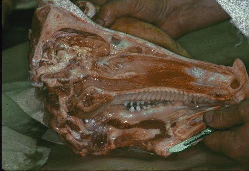 The nasal cavity of the skull (seen here dorsal to the roof of the moth in this split pig's head) contains the turbinate bones. Left and right turbinate bones have a shape that resembles a loosely rolled sheet of paper, which creates a large surface area for the nasal epithelium. However, in the view above, only the medial surface of the roll is apparent.
- The nasal cavity opens into the pharynx (shared with the alimentary canal), and then opens into the larynx.
- The larynx has a cartilagenous skeleton with muscles that support and stretch the vocal cords. In poultry, however, sound is produced by a separate organ, the syrinx, which is located farther down the respiratory system.
- The epiglottis is a spout-shaped cartilage that protects the entrance to the larynx.
- The larynx leads to the trachea or windpipe.
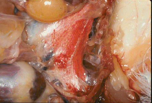
Trachea
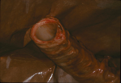
The trachea is a flexible tube held open by rings of cartilage, rather like a vacuum-cleaner hose, as shown above. If it was not held open, it would collapse when the animal tried to breath in. The continuity of each ring of cartilage is broken by a small dorsal gap. This image shows a transverse section through tracheal cartilage.
The trachea divides into two bronchi at a "Y" fork (Figure 1-15).
Bronchus
The bronchi connect with the right and left lungs, where they branch into progressively smaller ducts called bronchioles.
Epithelium
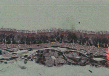
The trachea, bronchi and bronchioles are lined with ciliated epithelium and mucous glands, as seen in the image above which is a transverse section through the wall of the trachea. The cilia seen in a row across the top of the cells in tha image above (facing into the lumen of the trachea) are extremely fine whip-like hairs on the lumenal surfaces of cells. A complex system of mobile protein strands along the length of each cilium provides the motive power for movements that appear whip-like. Millions of cilia beat in a coordinated manner so that they can propel a continuous stream of mucus from the lungs to the nasal cavity. Thus, any small particles that have entered the lungs, despite the protective filtering of incoming air by the turbinate bones, can be removed.
Beef Lung
Gaseous exchange
- This occurs between inhaled air and the blood in the lungs, and takes place across the moist surfaces of ALVEOLI or alveolar sacs.
- In mammals, the alveoli are the final blind-ending branches of the air duct system.
- Beneath the moist epithelium which lines each alveolus is an extensive meshwork of lung capillaries.
- Oxygen is taken up by the blood in a loose combination with the hemoglobin of red blood cells or erythrocytes.
- There are three ways in which carbon dioxide may be carried in the blood; (1) in solution, (2) combined with blood proteins, or (3) as bicarbonate.
- Carbon dioxide is more soluble and diffuses faster than oxygen. The ratio of bicarbonate to carbonic acid determines the pH or acidity of the blood. This ratio is regulated by the rate of escape of carbon dioxide from the blood in the lungs: loss of carbon dioxide increases pH (decreases acidity).
- Gaseous exchange does not occur across the walls of the major air ducts that lead into the lungs. Thus, the last fraction of air that is inhaled becomes the first fraction to be exhaled, and the oxygen it contains is not utilized.
- Typical resting rates of respiration are 12 to 18 breaths per minute in cattle, 12 to 20 in sheep and 10 to 18 in pigs.The rate of respiration is controlled by the medulla oblongata in the posterior part of the brain. The medulla responds primarily to the pH and the carbon dioxide content of the blood; it increases the rate of respiration if the blood becomes acidic with a high level of carbon dioxide.

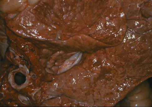
Pork Lung
Pleural membranes
When the lungs are removed from the body, slippery pleural membranes may be seen covering both the inner surface of the thoracic cavity and the lung surface. Pleural membranes prevent friction between the lungs and the body wall.
Inspiration and expiration
Caused by movements of the intercostal muscles, the ribs, the diaphragm and, sometimes, the abdominal muscles.
Diaphragm
The diaphragm resembles a strong drumskin that divides the thoracic and abdominal cavities, but it is thickened by muscle where it joins the body wall. In a dressed carcass, the muscular part of the diaphragm remains as a flap of muscle running diagonally across the inside of the ribcage.
By-Products
The proteins of the lungs (as well as those of the rumen and spleen), may be recovered by alkaline extraction followed by reacidification. Protein may be isolated as a powder or texturized to form fibers. Lungs also may be processed to isolate heparin, an anticoagulant for medical use.
Poultry
The respiratory system in poultry is quite different from that found in mammals (Figure 1-15). The lung is the red tissue in the image above.
- There is no diaphragm separating thoracic from abdominal cavities.
- Instead of being drawn into the lungs and then exhaled, air is drawn through the lungs and into air sacs outside the lungs. On exhalation, the air passes back through the lungs to the exterior.
- In poultry, therefore, the gaseous exchange between air and blood takes place as the air is moving through the lungs.
- The lungs of poultry are much smaller than those of mammals (relative to body size). Instead of occupying almost the whole of the thoracic cavity, they are located under the vertebral column where they are shaped to fit between the deep arches of the ribs where they meet the vertebral column.
The lungs of poultry are usually removed with a suction tube during commercial slaughter procedures whereas, in meat animals, the lungs are removed together with the trachea, bronchi and heart, as plucks.
The extensive system of AIR SACS in poultry extends between many of the viscera and even into certain bones.
- The interclavicular air sac is a single structure in the midline but the other air sacs are paired (right and left).
- The cervical extends towards the neck.
- The axillary is within the body at the junction with the wing.
- The anterior thoracic, posterior thoracic and abdominal sacs are in the body cavity.
- The humeral is located within the humerus as a branch of the axillary sac.
Air sacs have extremely thin walls and, when poultry are dissected, they should be identified while the viscera are in a relatively undisturbed condition.