6 Innervation of muscle
One of our final objectives is to
explain what regulates muscle growth and meat quality in our farm
animals. But we need to introduce a number of topics - all of which are
involved together. Meat is composed of myofibres (muscle fibres).
Myofibres come in a variety of types - differing in their pattern of
growth and contribution to meat quality. Myofibre types are controlled
by
- yes, you guessed correctly - the nervous system. In the
short term - second by second - the nervous system controls muscle
contraction. In the long term - week by week - the nervous system
controls how fast a myofibre contracts and how it obtains energy for
contraction. Control by the nervous system is - innervation.
6.1 Motor neurons
- A neuron is a nerve
cell.
- A motor neuron is a
nerve cell innervating myofibres.
- The cell
bodies of motor neurons innervating
meat carcass myofibres are located in the ventral horn of the spinal
cord.
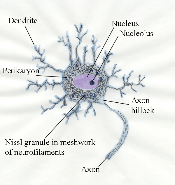
- The body
of the neuron is called the perikaryon.
- The
perikaryon has root-like projections called dendrites.
- A motor
neuron has one long axon
leading to the myofibres innervated.
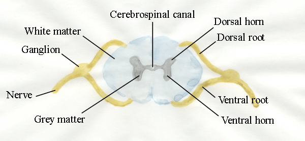 A transverse
section through the spinal cord.
- Basic
dyes for light microscopy show the
perikaryon is packed with Nissl
granules.
- Electron
microscopy reveals each granule is an aggregate of cisternae (membranous structures)
belonging to
the rough endoplasmic reticulum
(involved in protein synthesis).
- Colloidal
silver (as in a photographic emulsion) stains neurofilaments (diameter 10 nm)
which are bundled together to form neurofibrils.
- Neurofilaments
form a skeleton supporting the complex structure of a neuron.
- Silver
stains show the Nissl granules
occur in spaces surround by a tightly woven meshwork of neurofibrils.
- Interspersed
among the neurofibrils are a few 20 nm diameter neurotubules (microtubules).
- Small
mitochondria are found throughout the perikaryon and dendrites.
- The
single long axon linking each motor
neuron to its group of myofibres (motor
unit), usually departs from the
perikaryon at a small axon hillock devoid of Nissl granules.
- The axoplasm within the axon contains
tubules of endoplasmic reticulum, slender mitochondria,
neurotubules and neurofilaments.
- The
nucleus of the motor neuron has a
conspicuous nucleolus
(composed largely of RNA and showing the nucleus is no longer capable
of division by mitosis) .
6.2 Schwann cells & myelin sheaths
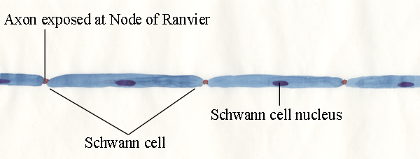
- Motor
axons in ventral nerve roots and
peripheral nerves are insulated by a myelin
sheath formed by Schwann cells.
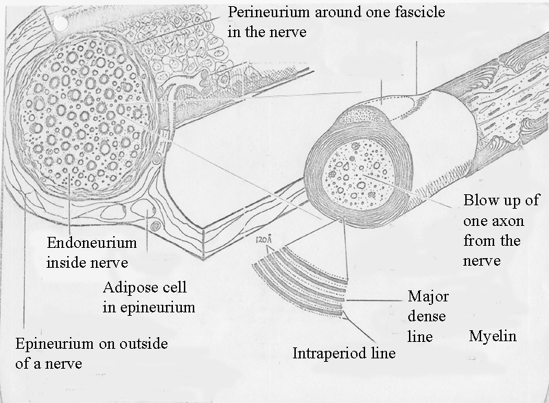
- The
Schwann cell is tightly rolled around the axon many times and
concentric
layers of Schwann cell membranes form a thick layer of insulation - myelin.
- In the
depth
of the insulating layer of myelin there are major dense lines (3-nm thick)
formed by the apposition
of the cytoplasmic faces of unit membranes, while faint intraperiod lines are
formed by apposition of outer faces.
- Major
dense lines are 12 nm apart.
- Schwann
cells migrate along axons and
become evenly distributed.
- Schwann
cells are separated by
nodes of Ranvier where the myelin leaves a narrow bare ring of axon.
- The
positions of the nodes of Ranvier are determined by the axon..
- Action
potentials (nerve impulses) jump from one node to another to increase
the velocity of neural conduction.
6.3 Nerves
-
A
nerve is composed of bundles of bundles of axons
surrounded by connective tissue.
Not a typo - I do mean bundles of bundles!
- Axons in
peripheral nerves are bound
together by an endoneurium of
fine collagen fibres, fibroblasts
(the cells that form the collagen fibres) and fixed macrophages (defensive
cells).
- Bundles
of axons are delineated by a more substantial perineurium
to form fascicles.
- Fascicles
are bound together by epineurium to form a
complete nerve with a blood supply and often with protective adipose
cells.
- The
smallest diameter axons (0.3 to 0.4 micrometres) in peripheral
nerves are not myelinated, and are mainly postganglionic sympathetic fibres
and
afferent fibres of the peripheral nervous system entering the dorsal
roots of
the spinal cord.
- The
smallest myelinated axons (up to 3 micrometres
diameter) are efferent preganglionic
fibres of the autonomic nervous system.
- Myelinated
sensory and motor axons range in diameter from 1 to 22
micrometres.
- In
nonmyelinated axons, conduction velocity is proportional to the
square root of axon diameter whereas, in myelinated axons, conduction
velocity
is proportional to diameter.
6.4
Motor unit
- One motor neuron innervates a group of myofibres - the
group is called a motor unit.
- All the
myoibres of a motor unit are within one muscle - but they
are scattered through the muscle.
- All the
myofibres in a motor unit contract at the same time when activated
by their motor neuron.
- The
contraction of motor unit is all or nothing - the
myoibres are
either relaxed, or contracting as hard as they can.
- Thus,
fine control of muscle activity is regulated by the number of motor
units contracted.
- Muscles
capable of precise contraction have many small motor units (really only
the extraocular muscles controlling the eyes of our meat animals).
- When one
motor unit is fatigued, another may replace it.
- Only
terrified animals can contract all the motor units in a muscle at once
- this can produced an astonishing amount of force. Even strong muscle
movements do not normally activate all motor units.
- When an
axon (inside a nerve) enters a muscle, it branches
to innervate all the scattered myofibres of its motor unit.
- In
small muscles, the
zone of myofibre innervation may be in an equatorial plane.
- In
most muscles
of meat animals the situation is more complex and, in pennate muscles (with an angular
arrangement of muscle fibres), even
short fasciculi in which all myofibres run from one end to the
other may
have two innervation bands.
-
This is advance warning - the
arrangement of myofibres in meat animals is complex and has
some important consequences when we study muscle growth in meat animals.
- The terminal
axon is the final axonal
branch innervating a myofibre.
- The
functional terminal innervation
ratio (FTIR) is the mean number of myofibres innervated by
the axonal
branches radiating from an intramuscular nerve.
- The
absolute terminal innervation ratio
(ATIR) gives the mean number of
neuromuscular junctions or motor end plates innervated by the axonal
branches
radiating from an intramuscular nerve.
- Both
FTIR and ATIR are increased when our meat animals have extra myoibres
(as in the case of double-muscled cattle).
6.5 Neuromuscular junctions
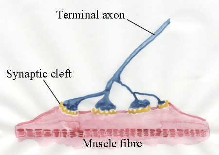
- The neuromuscular junction
is where a terminal axon innervates its myofibre.
- Neuromuscular junctions in farm mammals are compact in
structure and are called motor end
plates.
- At the
neuromuscular junction, the terminal
axon forms a terminal ramification
of small branches, often with bulbous ends.
- The
terminal ramification sits on a mound on the myofibre called the Doyère hillock.
- The
terminal ramification and the Doyère hillock
constitute a motor end plate.
- The
motor end plate is capped by perineural
epithelial cells (something like special Schwann cells).
- At
the point of contact between the terminal ramification and the myofibre
surface, the myoibre (covered by its basement membrane) is
corrugated to
form synaptic clefts.
- When an
action potential arrives at a motor end plate, synaptic vesicles in the
axoplasm fuse with the axonal
membrane, and each vesicle releases a quantal amount of acetylcholine into the
synaptic cleft.
- Acetylcholine
diffuses across the synapse, from the axonal
membrane to the myofibre membrane, and causes a change in the ionic
permeability of the postsynaptic area of the myofibre membrane.
- This
causes
the end plate potential to change.
- If
the change is of sufficient magnitude, it
initiates an action potential
on the surface of the myofibre, which then contracts.
- Acetylcholine
is rapidly destroyed by acetylcholinesterase
enabling the myofibre to
relax.
- In the
brief instant of one second, this whole sequence of events at the
neuromuscular junction may occur several hundred times.
- The growth of motor end
plates
is correlated with the growth of myofibres until motor end plates
reach a
certain size, after which, myoibre growth is no longer accompanied
by
motor end plate growth.
Further information
Structure and
Development of Meat Animals and Poultry. Chapter 6.
 A transverse
section through the spinal cord.
 A transverse
section through the spinal cord.


