27 Endocrine Control of Growth
27.1 Somatotropic hormone (STH)
- Years ago, we were all taught about growth hormone produced by the anterior pituitary gland.
It had a magic
control over body size - too little and an animal did not grow, too
much
and the animal became a giant. But then "growth hormone" was found to
have
many other properties - such as making energy available for strenuous
exercise.
Eventually, because of its other functions, "growth hormone" was
re-named somatotropic hormone (= somatotrophin) -
meaning it has a general
function on the body, including growth.
- In comparison with other body cells, skeletal myofibres are
unusual
because
they are multinucleated and because they derive new nuclei from the
mitosis
of satellite cells. Originally, STH was thought to be the main factor
responsible
for directly stimulating mitosis. Much of the information on STH levels
in meat animals supported this belief, because STH has a strong effect
on skeletal muscle.
- Injection of STH into meat animals usually causes increases in
live
weight
gain, feed conversion and leanness.
- Plasma STH often declines with age so it is correlated with
average
daily gain and feed efficiency.
- STH levels are particularly high at birth or hatching.
- A primary function of STH is to conserve muscle tissue at the
expense
of
fat. Thus, STH tends to act in the opposite direction to insulin,
This accounts for the increases in STH production found in
exercised
as well as starved animals - see why the name of the hormone had to be
changed?
- Anabolic steroids such as diethystilboestrol may
act
in ruminants
by increasing STH secretion, although this is not always the case in
all meat animals.
- Breeds of cattle with rapid early growth and a large mature size
may
secrete
more STH than small, slow-growing breeds. However, this is not always
the
case. Sometimes there may be no relationship between STH levels and
rate
of growth, or the relationship may be negative. Many of the
exceptions,
however, may be explained by the indirect action of STH in maximizing
lipid
utilization for energy requirements and in maintaining essential
protein
synthesis under unfavourable circumstances.
- Another factor to take into account is STH may be released in
surges
throughout
the day. Thus, single measurements may be of little value in estimating
the overall status of STH activity. Animals also show an enhanced
response
to exogenous STH if they are injected at intervals of several hours in
a manner that simulates the natural pattern of STH release.
Somatostatin is a peptide inhibiting the release of STH as well
as the release of insulin and other hormones affecting growth. It is
possible
to increase the growth rate of lambs by auto-immunization against
somatostatin.
The carcasses of treated lambs are heavier than normal but have normal
ratios of muscle to fat to bone. However, the initial
experiments
on the enhancement of growth by immunization against somatostatin were
conducted on unimproved breeds of sheep and the enhancement of
growth
in improved breeds is far less impressive.
27.2 Insulin-like growth factor (IGF)
Another name change! Back in the days of "growth hormone", it was
added to cultered muscle cells in anticipation of a surge of extra
mitotic
activity. But nothing happened. "Growth hormone" was then
tried
on many other types of tissue, and one did respond - liver. The
exciting
next experiment was - what would happen when "growth hormone" was added
to a culture of both types of tissue (muscle + liver). Yes, both types
of tissue now had a surge of mitotic activity in response to the added
hormone. What happens? The liver responds to the "growth hormone" by
producing
another hormone, and this other hormone is the one actually stimulating
mitosis in both types of tissue. At first, this new hormone was called
somatomedin. But then somatomedin was found to be like a very
potent
form of insulin, so the name was changed to insulin-like growth
factor
(IGF).
Sulphation factor
- The importance of STH as a primary growth
stimulant has been eclipsed by IGF.
- The restoration of STH to hypophysectomized animals
(unable to
produce
their own STH because of surgical removal of the anterior pituitary
gland)
causes a growth spurt in cartilagenous growth zones (from epiphyseal
plates).
- The growth of cartilage is conveniently monitored by the rate of
sulphate incorporation
into the matrix. Thus, we have a nice measure of growth.
- Even high levels of STH produce only slight increases in the rate
of
sulphate
incorporation. The action of STH is mediated by IGF from the liver in
response
to STH.
- In the oldest reports, therefore, IGF is called sulphation
factor.
IGF-1 and IGF-2
- Farm mammals have two forms of IGF, 1 and 2, both with
appropriate
receptors
and acting separately in myogenesis, whereas poultry may have only one
type of receptor.
- The distribution of receptors in muscle may be quite
complex. In
lambs, Type 1 and 2 receptors may differ between muscle and connective
tissue, depending on the animals' nutritional state.
- Hormonal IGF in the blood is complexed by binding proteins (IGFBPs),
of which several types are known.
- IGFBP modulates the function of IGF by inhibiting or stimulating
target
tissues.
- IGF-1 = somatomedin C.
- IGF-2 = somatomedin A.
Insulin-like properties
The effects of IGFs are widespread: they stimulate
(1) the transport of glucose,
(2) the uptake and incorporation of amino acids in muscle, and
(3) collagen synthesis .
- IGF is distinguished from insulin by not being blocked by
anti-insulin
serum, and by being present in hypophysectomized diabetic rats treated
with STH.
- A very
high concentration of insulin (relative to normally blood
levels)
stimulates differentiation in cultured premyoblasts. In this case, the
insulin appears to act as an analog of IGF.
- In contrast to this, IGF produces an equivalent stimulation of
premyoblast
growth and differentiation while at a normal low
physiological concentration.
- Porcine serum certainly contains something stimulating
myogenic
cells
in vitro, and these circulating factors are more effective when
isolated
from genetically lean pigs than from genetically obese pigs.
27.3 Control of protein synthesis in myofibres

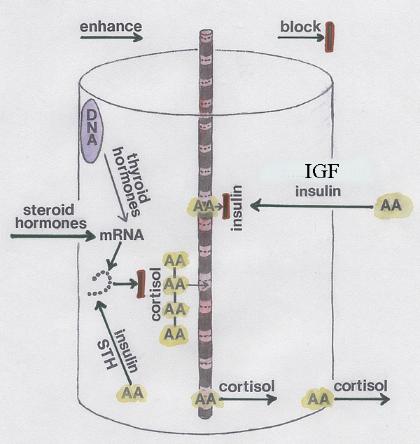
- The hollow cylinder represents a myofibre with just one of many
striated myofibrils shown down the axis.
- The arrows show
things with a positive effect (sometimes these enhance muscle growth -
sometimes they reduced muscle growth).
- The bar blocking the arrows shows things with a negative effect.
- Thyroid hormones increase messenger
RNA formation.
- Steroid hormones enhance messenger
RNA activity.
- Insulin and STH enhance the
transport of amino acid building blocks for the polyribosomal assembly
of proteins.
- Cortisol blocks the assembly of proteins (AA-AA-AA-AA etc).
- Insulin blocks proteolysis and the loss of already assembled
proteins.
- Cortisol accelerates the loss of already assembled proteins.
- Insulin blocks the release of already assembled proteins.
- Cortisol facilitates the release of amino acids from the myofibre.
- IGF and insulin increase the uptake of amino acids into the
myofibre.
27.4 Cortisol and animal stress
- Cortisol is a powerful glucocorticoid
hormone produced by the adrenal gland.
- It is based on cholesterol and its production is
regulated by pituitary ACTH
(adrenocorticotropic hormone) itself regulated by CRF (corticotropin
releasing factor).
- Cortisol has a negative feedback by inhibiting ACTH
and CRF production.
- Most cortisol in blood plasma is tightly bound to corticosteroid binding globulin
(CBG).
- Cortisol acts on intracellular
receptors.
- It has many different effects on the body, including
glucose metabolism, functioning of the immune system,function, blood
vessel diameter and bone metabolism.
- Cortisol is
produced by stressed animals in order to help cope with the
stress by increasing blood pressure and blood glucose (useful if the
animal has to fight or run).
- But farm animals should not be stressed if they are being
properly reared.
- You can see in the diagram above how cortisol will
prevent muscle growth.
Further information
Structure and Development of Meat
Animals and Poultry. Pages 476-483.
