26 Bone
Growth and Calcium Metabolism
26.1
Introduction
Bone is
important for meat animal production in several ways.
- Longitudinal growth of muscles accounts for much of the meat we
produce. The longitudinal growth of muscles is limited by the
longitudinal growth of bone. When we select animals to produce
more meat, we often increase the length of their bones. Thus,
frame size (the maximum size of the adult skeleton) is related to
efficient meat production.
- Bones store calcium. Bone calcium is used for milk
production and the formation of egg shells in poultry.
- When we grade beef, we need to know the approximate age of the
animal. If the head remained on the commercial beef carcass, we might
get this information from the state of the dentition. Without this, we
use the degree of ossification of the carcass. The general
principle is bones are first formed from cartilage - then progressively
ossified. Thus, the extent of ossification tells us the approximate age
of the animal.
26.2 Cartilage
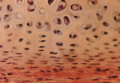
-
Cartilage cells or chondrocytes
occupy lacunae in a
stiff flexible matrix formed
from collagen
fibres in a proteoglycan
ground substance. Imagine the lacuna (plural = lacunae) as a cave in
the
matrix. The chondrocyte fills the lacuna.
- Hyaline cartilage has a white
translucent appearance and occurs on the smooth surfaces of joints. In
the
larynx, trachea and bronchi, hyaline cartilage forms the rings that
hold these
air ducts open during respiration. Flexible units of the skeleton, such
as the
dorsal part of the scapula and the linkages between the sternum and the
posterior ribs, also are formed from
hyaline cartilage.
- Most of the
bones of the carcass are initiated prenatally as
cartilagenous models. Complete ossification
is a slow process, and the bones of young meat animals are more
flexible than
those of adults. The state of ossification is a useful clue to animal
age in
carcass grading.
- Chondrocytes
are derived from
mesenchymal cells and are initially capable of both mitosis and matrix
formation.
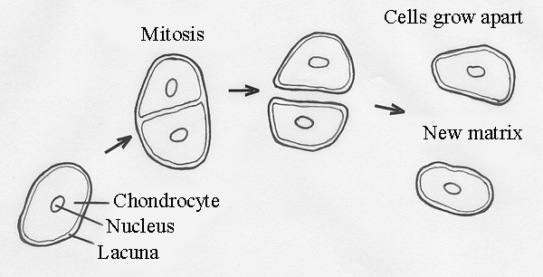
- Clusters of
related cells are pushed apart by their new matrix in a
process called interstitial growth.
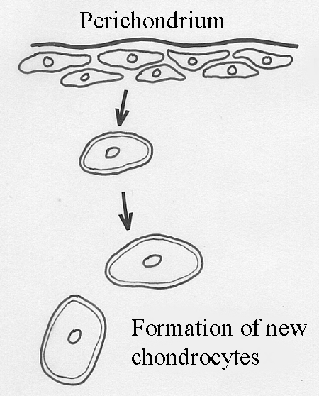
- Cartilaginous
models of
prenatal bones are covered by a membrane known as the perichondrium. Inner
perichondrial cells differentiate into chondrocytes. Thus, in
addition to
interstitial growth, new cells and matrix may be added superficially in
a
process known as appositional growth.
- Cartilage may
acquire
numerous elastic or collagen fibres to become elastic cartilage or
fibrocartilage, respectively. The dominant type of collagen in hyaline
cartilage is Type II and accounts for 50 to 70% of dry weight.
Elastic cartilage is found in parts of the body such as the ears
and muzzle.
26.3 Bone
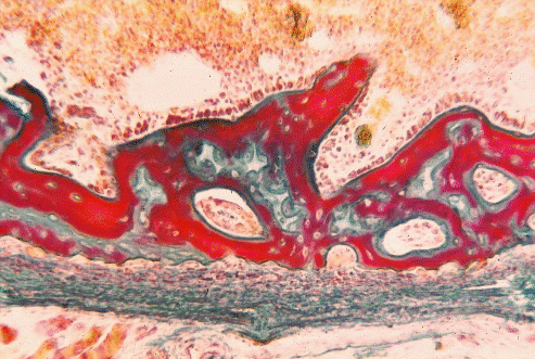
In the section of bone shown above, the marrow cavity is at the top,
the calcified matrix is stained red, and the periostium runs acroos the
bottom.
- Oxygen,
nutrients and waste
products may travel to and from the chondrocytes in cartilage by
diffusion
through the surrounding matrix but, when the matrix becomes ossified by
the
deposition of submicroscopic hydroxyapatite
crystals, diffusion is greatly
reduced.
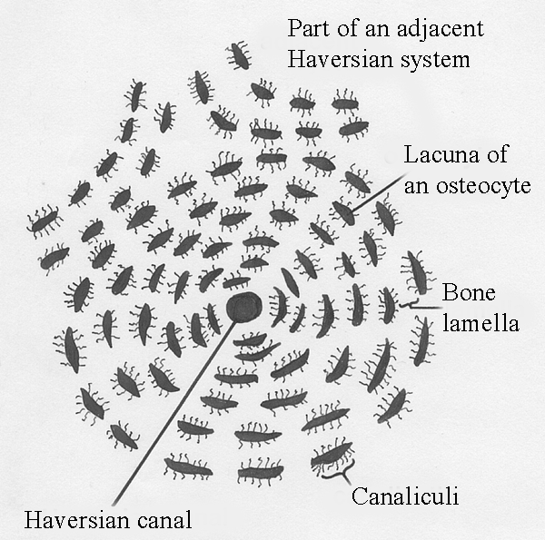
- Bone cells, osteocytes, can only survive if they
develop long cytoplasmic
extensions radiating from their lacuna to regions where exchange by
diffusion
can take place. These cytoplasmic extensions run through fine tubes or canaliculi in the ossified
matrix, but are limited in length. Consequently,
large numbers of blood vessels permeate the matrix of bone.
- Most of these
blood
vessels run longitudinally
through the bone in large haversian
canals
surrounded by concentric rings of osteocytes and bone lamellae.
- Bones
are covered by a connective tissue membrane called the periosteum.
The prenatal
formation of bone
is initiated by either of two basically different processes, either
intramembranous or endochondral ossification.
Intramembramous ossification
is typical of the bones forming the vault of the skull, and occurs when
sheets of connective tissue produce osteoblasts
which then initiate centres of
ossification. Endochondral ossification is more common, and is the
process by
which cartilagenous models become ossified to form the bones of a
commercial
meat carcass.
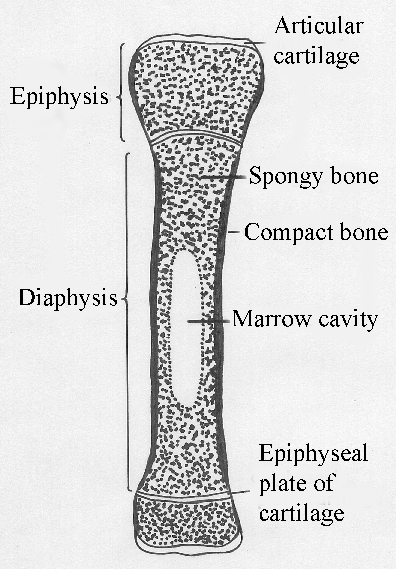
- The
internal structure of
carcass bones becomes visible when they are split longitudinally on a
band saw.
- The shaft
of a bone is called the diaphysis and
often includes a marrow cavity with
a
variable amount of fat.
- The
knob at each end of a bone is called the epiphysis.
- Between
the diaphysis and
each epiphysis is a cartilagenous growth plate called the epiphyseal plate.
- In
a young animal, the chondrocytes of the epiphyseal plate are constantly
dividing to form new matrix. However, on each face of the plate,
cartilage is
continuously resorbed and is replaced by bone. Hence, the
thickness of the epiphyseal plate tends to remain constant in growing
animals.
- This
process allows a bone to grow longitudinally without disrupting the
articular surface on the epiphysis.
- The rate
of the longitudinal growth of
bones is the product of two factors; (1) the rate of production of new
cells,
and (2) the size that cells reach before they degenerate at the point
of
ossification.
- The
strength and thickness of
epiphyseal plates is modified by sex hormones. At puberty,
chondrocyte growth slows down and fails to keep pace with ossification
on the
surface of the epiphyseal plate. Thus, epiphyseal
plates are lost in mature
animals, and the epiphyses become firmly ossified to their diaphyses.
- However,
factors regulating the closure of the epiphyseal plate in meat animals
and, hence, the
frame size of the animal, are poorly understand. Although regulation is
likely
to be an interaction between animal age and circulating hormones, there
are no
obvious hormonal changes when the plate closes and
nutrition and management have a minimal effect. If
whethers are implanted with estradiol the ossification of growth plates
is
accelerated.
- Bone growth
in mature animals is restricted
to the girth or thickness of the bone, and occurs by recruitment of
periosteal
cells to become osteoblasts in response to loading by body mass<>.
26.4 Bone calcium
- Radioactive
calcium (45Ca) is used to study the uptake of calcium by the
skeleton.
- There is a
continuous exchange of calcium between the body fluids (mostly plasma)
and
approximately 1% of the total bone matrix which has`direct access to
the circulating
fluid.
- Growth by
accretion results from small net gains to the matrix.
- When
radioactive calcium is injected into a growing animal, the isotope
is incorporated into new bone, and the concentration of isotope in the
plasma
declines.
- Both calcium
exchange and calcium accretion are
more rapid in the epiphysis than in the diaphysis.
- Calcium,
phosphate and
hydroxyl ions are obtained from the extracellular fluid during bone
formation.
The first stage in ossification is the deposition of a crystal of calcium
phosphate, then calcium phosphate is converted to hydroxyapatite.
- The
supply of calcium and
phosphate from the blood is affected by vitamin D. Vitamin D is obtained
from
the diet or by exposure to
ultraviolet light. It is hydroxylated
in the liver and then converted to the hormonal form
(1,25‑dihydroxycholecalciferol)
in the kidney. The hormonal form causes the
intestine to increase its absorption of calcium and phosphate.
- The
resorption of bone enables
bone remodelling in response to local stresses and is coupled with the
maintenance of blood calcium and phosphorus levels.
- The organic
components of bone are degraded by the lysosomal enzymes of osteoclasts, a
process requiring vitamin A.
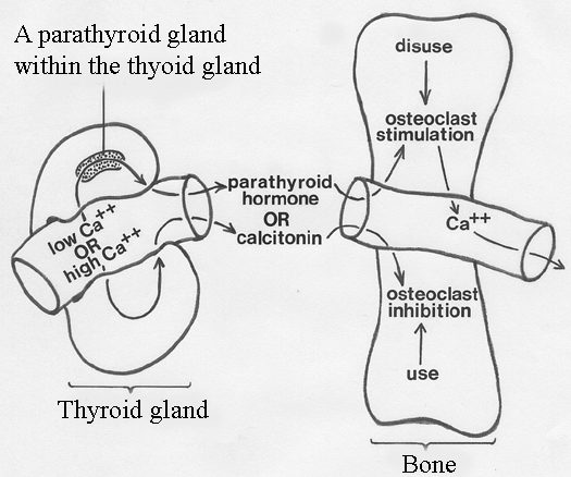
- The
solubilization of hydroxyapatite in
response to parathyroid hormone
(from the parathroid glands) is achieved by a combination of low pH
(due to anaerobic glycolysis) and chelation. Osteclasts (cells which
erode bone) then release calcium into the blood stream.
- Osteoclasts
are inhibited by calcitonin
(from the thyroid gland) so that calcium tends to be stored in bone and
the level in blood is decreased.
- Genetic
defects in
calcification and bone formation may occur in cattle. Typical signs are
lack of
calcification of the teeth and hypermobility of joints.
- In milk fever
of cows (parturient paresis),
paralysis and unconciousness
may occur during parturition or early lactation. The condition is a
result of
drastically lowered plasma calcium and inorganic phosphorus levels, and
may be
treated by the injection of calcium
borogluconate.
26.5 Control
of bone growth
-
Carcasses from young animals
have a relatively high bone content because the skeleton is well
developed at
birth.
- As an animal
grows to market weight, its proportion of bone decreases on
a relative basis, because of the growth of muscle and fat.
- The long‑term
control of bone growth is superimposed on the short‑term regulation of
bone
metabolism that occurs in response to changes in blood calcium levels
or to remodelling in response to local functional demands.
- A
number of hormones exert
secondary effects on skeletal development. Thyroxine, insulin, somatotrophin
and gonadal hormones tend to be anabolic. Estrogens may inhibit
resorption of
bone. Adrenal corticosteroids stimulate resorption of bone and inhibit
the
formation of new bone.
- In cattle, castration
delays the completion of growth in
epiphyseal plates. This is most noticeable in the distal
bones of the limbs and enables the continued longitudinal growth of the
legs.
In the vertebral column, however, castration reduces bone growth..
- Removal of
the ovaries from heifers causes an increase in the longitudinal growth
of distal bone.
- Bone growth
is regulated by transforming growth factor (TGF‑ß) and insulin-like growth
factor I (IGF I).
How do bones adapt
to patterns of use?
One hypothesis is loads frequently placed
on a region of bone
cause the transduction of mechanical energy to electrical energy by a
piezoelectric effect. In a
frequently loaded and negatively charged region,
growth is stimulated. In
electrical fields, osteoclasts migrate towards the positive electrode
while osteoblasts migrate
towards the negative electrode. In an unloaded and
positively charged region, resorption is stimulated.
Why do bones from
older animals have more knobs and wrinkles?
Because
most farm animals are slaughtered in a fairly
immature condition, the relationship between muscles and the bony
processes they pull on may not be immediately obvious. But knobs
and wrinkles on
bone surfaces become more conspicuous with age, and they are readily
seen in
the carcasses of old bulls. One possible relationship between muscle
activity
and bone growth may be muscle contraction - by stopping or slowing the
venous blood flow, it may stimulate bone growth. Alternatively, by
pulling on the
periosteum, the effect of muscle activity may be mediated by connective
tissue.
The importance of local factors is seen in bone transplants, where
growth of
the transplant almost immediately becomes regulated by the new local
conditions.
- Breeds of
cattle with a large
mature size usually produce lean meat at a faster rate than early
maturing
traditional beef breeds with a relatively small adult size.
- Differences
in
adult size are produced by differences in skeletal growth.
- Meat quality
is involved because
large‑framed animals produce leaner meat during their production life
span.
- Large‑framed
breeds mature late and have a later cessation of linear skeletal growth
at their epiphyseal plates.
- The time of
maturation is related to the
distribution and amount of adipose tissue in the carcass, particularly
marbling
fat, and could well be regulated by leptin,
a hormone secreted by fat and
involved with ingestive behavior and energy balance,
mediated via a receptor in the brain. Thus, because leptin
normally inhibits glycogen synthesis in muscle, decreased
fat deposition may lead to abnormal glycogen accumulation, which
predisposes
pigs to abnormally rapid or extensive postmortem glycolysis, and to the
formation of PSE pork.
- Differential
bone growth
between large and small breeds of cattle is usually established prior
to a
slaughter weight of 500 kg in males.
- Comparing
present day cattle with those born 20
years ago, the faster growing modern animalsare longer in body,
but not necessarily taller than their predecessors.
- Pelvic
dimensions in cows of
different breeds are related to the incidence of difficult calving or
dystocia.
- Growth
promotants may
have little or no effect on skeletal development,
SOME HISTORY. In
the
early 1950s, attempts were made to use measurements of isolated carcass
bones
such as the cannon bone to predict the muscle
to bone ratios of carcasses. Although
the method worked satisfactorily when applied to a wide range of
dissimilar
carcasses, it was of little practical value when applied to more
uniform
commercial carcasses. Muscle to bone ratios improve as animals grow
older or
fatter, since longitudinal bone growth slows down in older animals and
muscles
start to accumulate appreciable amounts of intramuscular fat. Animal
age is the
dominant factor that determines muscle to bone ratios.
Some years
ago, the desire to produce small compact
animals with
bulging muscles favoured the survival of dwarf animals with impaired
longitudinal bone growth. Although mildly affected animals looked very
muscular, severely affected animals became increasingly common and were
poorly suited for beef production. Dwarfism
from impaired longitudinal growth
of bones is a recessive trait that affects males more strongly than
females.
Further information
Structure and Development of Meat
Animals and Poultry. Pages 96-106.


