- In a live animal, the lumbar vertebrae act like a
suspension bridge to support the weight of the abdomen. The lumbar
vertebrae have flat, wing-like transverse processes that broaden
the abdominal cavity dorsally to provide a strong attachment for
the muscles of the abdominal wall that carry the weight of the
viscera.
-
Plan of
kumbar region in hanging beef carcass showing: 1, ilium; 2, sacrum;3,
last lumbar vertebra;4, first lumbar vertebra; and 5, last rib.
- The propulsive thrust generated by the hindlimb during
locomotion is transmitted to the sacral vertebrae by the pelvis. To
strengthen the sacral vertebrae, they are fused together to form
the sacrum.
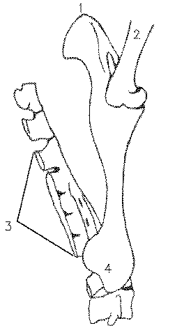 Plan of the sacral region in a
hanging beef carcass showing:1, ischium; 2, femur;3, sacrum; and 4,
ilium.
Fusion of the sacral vertebrae to form the sacrum is incomplete
in young animals and provides an important
clue to animal age in the dressed carcass.
- The pelvis is formed by three bones on each side. The most
anterior bone on each side is the ilium. The shaft of the ilium
expands anteriorly to form a flat wing attached to the sacrum. This
joint is called the slip joint. When seen in a sirloin steak, the
ilium may appear either as a small round bone or a large flat bone.
The anterior edges of the ilia form the hooks of the live animal.
The most posterior bone of the pelvis on each side is the
ischium.
- The pelvis and the sacrum form a ring of bone completed
ventrally by the pubes. The left pubis is separated from the right
pubis by fibrocartilage which, at parturition, may soften to allow
movement between the bones of the pelvis. The pubes are separated
when carcasses are split into left and right sides in the
abattoir.
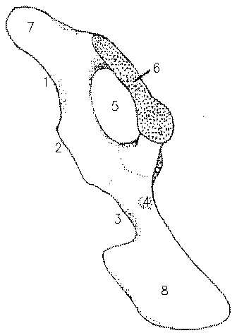 Plan of the
pelvis in a hanging beef carcass showing:1, lesser sciatic notch; 2,
ischiatic spine; 3, greater sciatic notch; 4, psoas tubercle; 5,
obturator foramen; 6, symphysis pubis;7, ischium; and 8, ilium.
- The pubic bone exposed on a carcass is called the aitch bone.
The aitch bone is curved in steer and bull carcasses, is
moderately curved in heifers, but is straight in cow carcasses.
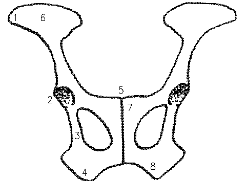
Another
plan of the both sides of the pelvis in a hanging carcass showing: 1,
tiber coxae; 2, acetabulum; 3, acetabular ramus of ischium; 4, tuber
ischii; 5, symphysis pubis; 6, ilium; 7, pibis; and 8, ischium.
- Only two caudal or coccygeal tail vertebrae are left on a
commercial beef carcass.
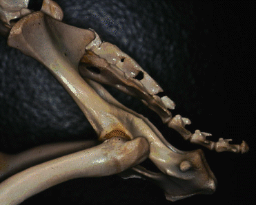 Sheep pelvis
 Sheep pelvis
 Plan of the sacral region in a
hanging beef carcass showing:1, ischium; 2, femur;3, sacrum; and 4,
ilium.
 Plan of the
pelvis in a hanging beef carcass showing:1, lesser sciatic notch; 2,
ischiatic spine; 3, greater sciatic notch; 4, psoas tubercle; 5,
obturator foramen; 6, symphysis pubis;7, ischium; and 8, ilium.
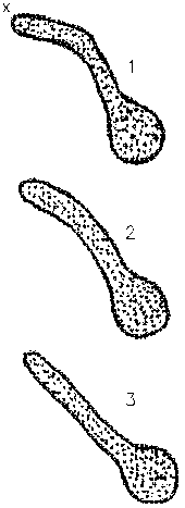 The shape
of the symphysis pubis seen on a side of beef is a useful guide to the
sex of the carcass: 1, steer; 2, heifer; and 3, cow, where x shows the
position of the pizzle eye.