LAB 8.2 Forequarter muscles
PLAY VIDEO
Neck muscles
The neck muscles
produce tough meat
because
of their high connective tissue content. Like other tough muscles in
the
carcass, they are usually removed from their bones (cervical vertebrae)
and are
used as stewing meat or ground beef. Since the anatomy of the neck
muscles is
rather complex, and since their individual identity is of no
consequence once
the carcass is subdivided, they need not be considered in detail here.
The only
muscle worthy of passing note, however, is a strap‑like muscle called
the
sternomandibularis. This muscle
forms the superficial part of a compound muscle
in the ventral throat region running from the head to the sternum.
Because this
muscle is severed when the head of a beef carcass is removed at
slaughter,
samples may be obtained immediately after slaughter without damaging a
commercial carcass.
SHOULDER
MUSCLES .
Shoulder muscles are
intermediate in
their
level of toughness and are usually completely cooked in order to make
them
tender. An easily recognized group of muscles and bones enables meat
from the
shoulder or chuck region to be readily identified. The supraspinatus is dorsal
to the spine or ridge on the scapula, while the infraspinatus is ventral to the
scapular spine. The trapezius
is located superficially between the left and
right scapular blades. The rhomboideus
is ventral to the trapezius.
Figuratively speaking, if one were to stab a standing animal between
its
shoulder blades, the knife would pass through the trapezius first and
then
through the rhomboideus. The subscapularis
is located on the flat medial face of
the scapula, towards the ribs. The biceps
brachii is anterior to the humerus in
an equivalent position to the biceps muscle in the human arm. The
adjective
brachii is needed to indicate the biceps muscle of the arm, since there
is
another biceps muscle, the biceps femoris, located in the hindlimb. The
triceps
brachii is a large muscle located in the triangular area
bounded by the ventral
edge of the scapula and the posterior edge of the humerus. Its name,
tri‑ceps
or three‑heads, indicates that this large triangular muscle is
subdivided.
Thus, when seen in a cut of meat from the shoulder, the triceps brachii
may
look like more than one muscle.
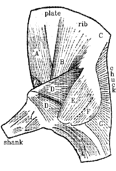
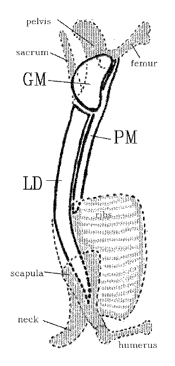
In the view of the superficial muscles (left diagram above) A is the
pectoralis, B is latissimus dorsi, C is the position of the longissimus
dorsi, D is triceps brachii, E is infraspinatus and G is trapezius. The
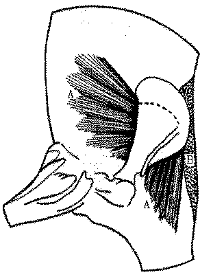
deep dissection (right diagram above) shows serratus ventralis (A) and
rhomboideus (B).
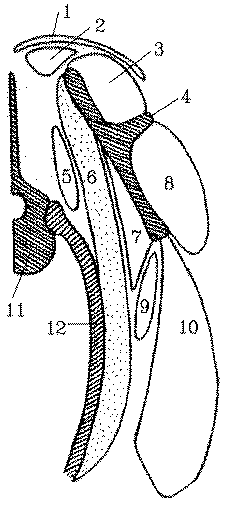
The above diagram is a section through the shoulder to show trapezius
(1), rhomboideus (2), supraspinatus (3), the spine of the scapula (4),
longissimus dorsi (5), serratus ventralis (6), suscapularis (7),
infraspinatus (8), an edge of latissimus dorsi (9), triceps
brachii (10), a thoracic vertebra (11) and a rib (12).
Distal
muscles of the limbs
The distal muscles of
the limbs produce
tough meat because of their high content of connective tissue. In the
distal
regions of the forelimb (shank) and hindlimb (leg) are groups of
fusiform
(cigar‑shaped) muscles with long tendons which extend distally towards
the toes
or phalanges. If these muscles are cut open longitudinally, it may be
seen that
most of them contain internal tendons that fan outwards for the
attachment of
short bundles of muscle fibers. This feather‑like arrangement of muscle
fiber
bundles is called a pennate structure. In meat animals, the meat
derived from
pennate muscles is quite tough, because of their high connective tissue
content.
Most of the distal
muscles located
anteriorly in the limb are extensors and, when they contract in the
living
animal, they cause the toes to move forwards, as in the start of a new
stride.
Most of the muscles located posteriorly in the distal part of the limb
are
flexors which bend the limb during locomotion. In beef and lamb
carcasses,
tendons from distal muscles pass down the length of the cannon bone and
are
kept in place by ligamentous rings. Thus, the extremities of a limb are
moved
by remote control. Beef cannon bones are discarded in the abattoir
because they
have virtually no meat on them. Flexors and extensors from the distal
parts of
the limbs are difficult to identify individually once they have been
removed
from the skeleton.
Muscles
of the ribcage
Located between
the scapula and
the
ribcage are several muscles that hold the forelimb onto the body. Like
other
shoulder muscles around the scapula, most of these muscles are
intermediate in
their level of toughness. The serratus
ventralis is a large fan‑like muscle
that radiates from the medial face of the scapula and attaches to the
lateral
surfaces of the ribs. The serratus ventralis is the major
component of a muscular sling that suspends the thorax of an animal
from
between its forelimbs. This muscular suspension system has no
counterpart in
the hindlimb since the pelvis is fused to the vertebral column. The
muscular
sling that holds the forelimb onto the body serves as a shock absorber
during
locomotion.
The
longissimus dorsi is an extremely
important muscle. It forms the eye of meat seen when chops and steaks
are cut
from the posterior rib region and loin. The naming of
this muscle is rather a problem since it is really a compound muscle
composed
of many subunits. Each subunit acts
over the length of several vertebrae and helps to flex the vertebral
column.
The longissimus dorsi also is involved in respiratory movements, as
well as
helping to move the neck. Because of its compound structure, the
longissimus
dorsi may be called by an alternative name, longissimi thoracis et
lumborum. In
agricultural journals the longissimus dorsi is often simply called the
eye‑muscle
or longissimus muscle. The muscle fiber bundles of the longissimus
dorsi are
arranged at an acute angle to the vertebral column. The cross sectional
area of
the longissimus dorsi increases towards the posterior part of the
ribcage, but
it has an approximately constant cross sectional area through the loin.
Beef
carcasses are usually split into forequarters and hindquarters between
ribs 12
and 13. The area of the longissimus dorsi seen at this point is often
measured
or examined in order to assess the amount of meat in a carcass. This
may be a
useful guide to muscularity when comparing animals with a similar
carcass
length. However, in the comparison of long carcasses with short
carcasses, a
smaller cross sectional area does not necessarily indicate a smaller
muscle
mass, since the mass is spread over a greater length.
The
pectoralis muscle is located over the
sternum in the brisket, and it extends posteriorly into the plate. The
pectoralis
is composed of deep and superficial layers. The intercostal muscles are
located
between adjacent ribs in the wall of the chest, and there are two
layers in the
depth of the muscle. Intercostal muscles make an important contribution
to the
meat content of North American pork spare ribs.
Loin
muscles .
The loin muscles give
rise to tender
meat
with a desireable taste, and they command a high price when presented
for sale
as steaks or chops. The longissimus dorsi extends posteriorly from the
rib
region, it runs through the loin, and most of the muscle terminates on
the
anterior face of the ilium. Thus, the longissimus dorsi is seen in cuts
of meat
taken through the ribs and loin, but not in cuts of meat such as
sirloin steaks
that are posterior to the anterior face of the ilium. The longissimus
dorsi is
dorsal to the transverse processes of the lumbar vertebrae, and it is
dorsal to
the ribs in the thoracic region. For most of the length of the ribcage,
there
are no major muscles immediately ventral to the heads of the ribs.
Thus, in a
rib steak, there is only one main eye of meat, and that is the
longissimus
dorsi dorsal to the ribs. However, in the loin, there are muscles both
above
and below the level of the transverse processes of the lumbar
vertebrae. The
dorsal muscle above the transverse processes is the longissimus dorsi.
The
ventral muscle below the transverse processes is the psoas major or filet
muscle. The psoas major originates ventrally to the last couple of ribs
in the
ribcage. The cross sectional area of the psoas major increases towards
the
sirloin. Medial to the psoas major, almost under the centra of the
vertebrae,
is a smaller psoas muscle called the psoas minor. The letter P in the
word
psoas is silent when the word is spoken.
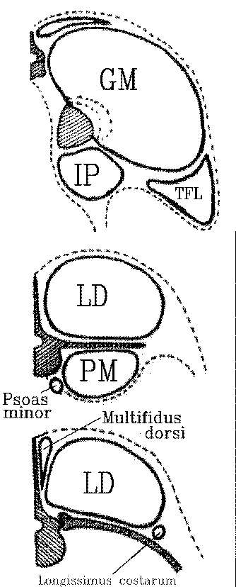
Immediately lateral
to the dorsal spines
of
the vertebrae (medial to the longissimus dorsi) are some small multifidus dorsi
muscles. The longissimus costarum
is a relatively small rope‑like muscle,
dorsal to the ribs. It appears as a small eye of meat at the separation
between
the forequarter and the hindquarter. The multifidus dorsi and the
longissimus
costarum have little commercial significance, since they are such small
muscles, but they may create a problem in the measurement of rib‑eye
areas
since they might be accidently included with the longissimus dorsi.
IThese are shown in the bottom part of the diagram below (we will leave the top two parts of the
diagram - from the hindquarter - for LAB 9).