LAB 8.1 Forequarter skeleton
PLAY VIDEO
Skeleton of the neck
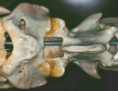 Atlas (on left) and axis of
sheep.
- The vertebral column or backbone is the main axis of
the skeleton and it protects the spinal cord. The spinal cord is
located in a neural canal formed by a long series of neural
arches, each contributed by a different vertebra.
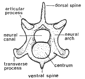
- The neural arch of each vertebra is supported on the body or
centrum of the vertebra. In some types of vertebrae, the neural
arch extends dorsally as a prominent spine that may be called a
dorsal spine, a neural spine or a spinous process.
- Where movement between vertebrae is possible, the centra are
separated by cartilaginous intervertebral discs. In mammals,
the anterior and posterior faces of the centra are almost flat.
- The names and numbers of the different types of vertebrae in
meat animals are variable.
NAME--------REGION---------BEEF----------PORK------------LAMB
Cervical------Neck-------------7--------------7----------------7
Thoracic-----Ribcage-----------13------------13 to
17-----------13 to 14
Lumbar-------Loin-------------6-------------5 to 7------------6
to 7
Sacral
------Sirloin-------------5-------------4-----------------4
Caudal-------Tail------------18 to 20----------20 to
23----------16 to 18
�
- Meat animals, like most other living mammals, usually have
seven cervical vertebrae in the neck region. However, sheep
sometimes have only six cervical vertebrae, and as few as five
cervical vertebrae have been reported in pigs.
- In cattle and sheep, but to a lesser extent in pigs, the neck
is very mobile, and the cervical vertebrae have a series of
interlocking articular and tranverse processes that limit
excessive bending of the neck to protect the spinal cord.
- The first cervical vertebra, the atlas, articulates with the
skull and is greatly modified in shape to form a joint that enables
the animal to nod its head up and down.
- Rotation or twisting of the head occurs from the joint between
the atlas and the next cervical vertebra, the axis.
- The ligamentum nuchae is a very strong elastic ligament in the
dorsal midline of the neck, and it relieves the animal of the
weight of its head. Were it not for the ligamentum nuchae, the head
of the standing animal would droop between its forelimbs.
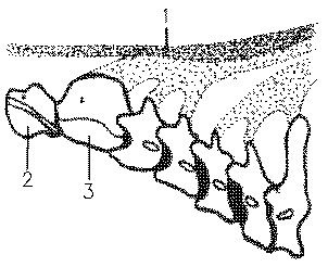 Plan of neck in beef, showing:1,
ligamentum nuch; 2, atlas; and 3, axis.
The ligamentum nuchae is pale yellow
 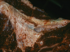 with a thick cord-like or
funicular part and a flat sheet-like or lamellar part. Once the
head is removed at slaughter, the elasticity of the ligamentum
nuchae causes the neck of the carcass to curve dorsally.
- Beef and pork carcasses usually are split into right and left
sides soon after slaughter and the series of vertebral centra that
are now exposed is called the chine bone.
Ribcage
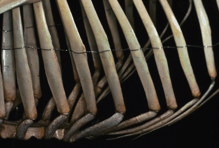 Sheep
Sheep
- The number of vertebrae in pork carcasses is rather
variable, particularly in certain breeds. The heritability of the
number of vertebrae is about 0.74. Each extra vertebra adds about
15 mm to the length of the carcasses at slaughter weight.
- The number of thoracic vertebrae, each bearing left and right
ribs, ranges from 13 to 17. Breeds with a large size when mature
and with heavy bone development tend to have more thoracic
vertebrae than lighter breeds. Sometimes the ribs on extra thoracic
vertebrae are only partially formed, but usually they are
complete.
- The minimum number of lumbar vertebrae is generally found in
cacasses with the maximum number of thoracic vertebrae. However,
the variability of vertebral numbers frequently leads to an
increase in the total number of vertebrae, so that the phenomenon
is not due simply to the substitution of one type of vertebra for
another.
- Lamb carcasses usually have either 13 or 14 thoracic vertebrae
and a corresponding number of pairs of ribs. Experimental studies
suggest that the number of vertebrae in an animal is determined by
the number of somites that develop along the length of the spinal
cord. By definition, in mammals, the vertebrae that bear ribs are
identified as thoracic vertebrae. In the embryo there are
ossification centers on each side of the developing vertebrae. In
vertebrae that do not normally develop ribs, these lateral
ossification centers contribute their bone tissue to the centra of
adjacent vertebrae. In the thoracic vertebrae, however, this
laterally derived bone tissue remains separate from the centra and
forms the ribs. Thus, the numbers of pairs of ribs and the numbers
of thoracic vertebrae are determined by the developmental mechanism
that controls the fate of the tissue which is derived from the
lateral ossification centers.
- The cage formed by thoracic vertebrae, ribs and sternum
is an essential component of the respiratory system. Thoracic
vertebrae are distinguished by their tall dorsal spines, many of
which point towards the hindquarter and are known as the feather
bones.
- The ribs are joined to the vertebral column dorsally so
that the head of each rib articulates with the bodies of two
adjacent vertebrae.
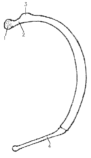 Rib showing : 1, head; 2, neck;
3, tubercle; and 4, costal cartilage.Each rib has a tubercle
that articulates
with the transverse process of the more posterior of its two
vertebrae. Ventrally, the anterior ribs articulate with the sternum
and are termed sternal ribs. The more posterior ribs are
called asternal ribs and they only connect to the sternum
indirectly via costal cartilages. The most posterior ribs have only
small costal cartilages that do not reach all the way to the
sternum. Some of the costal cartilages are very hard and may appear
more like bones than typical cartilage.
- The sternum is formed by a number of closely joined bones, the
sternebrae. When split through the midline, the interior
structure of the sternebrae resembles that found in the centra of
the vertebrae. But in an isolated cut of meat, the distinction
between sternebrae and vertebral centra may be made by the presence
or absence of a neural canal. A neural canal is seen in the
vertebrae of carcasses that have been symmetrically separated into
right and left sides.
- The structure of the ribcage is rather variable in lamb
carcasses. Carcasses have been found with as few as 12 ribs on one
side, and left and right sides of the ribcage may differ in their
number of ribs. In lambs, rib length is determined mainly by age
while the plane of nutrition determines rib thickness.
-----------------------BEEF---------PORK------------LAMB
Total pairs of ribs-------------13----------13 to 17-----------13 to
14
Pairs of sternal ribs-------------8-----------7-----------------8
Pairs of asternal ribs------------5-----------7 to 8-------------5
to 6
Number of sternebrae-----------7-------------6--------------6 to 7
Scapula
- The most proximal bone of the forelimb is the blade bone or
scapula.
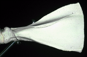 This
is the shape of the blade bone in beef.
The scapula is not fused to the vertebral column (like the
pelvis in the
hindlimb), and this allows muscles that hold the scapula to the
ribcage to function as shock absorbers during locomotion.
The scapula has a distal socket joint for the next bone in the
forelimb, the humerus. This socket joint is called the glenoid
cavity . The glenoid cavity is wide and shallow, unlike the ball
and socket joint in the hindlimb which is narrow and deep. Along
the dorsal edge of the scapula, the bone merges with flexible
hyaline cartilage. On the lateral face of the scapula is a
prominent ridge of bone called the spine of the scapula. In beef
carcasses, the scapular spine is extended distally as a prominent
acromion process.
Humerus
- Proceeding distally down the forelimb, the bone that
articulates with the scapula is the humerus.
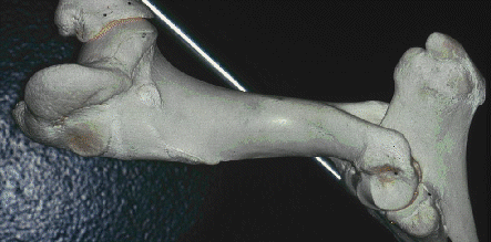 Proximally, the humerus has a relatively flat knob or head to fit
into the glenoid cavity of the scapula. Two well defined
condyles on the distal end of the humerus contribute to the hinge
joint at the elbow.
Radius & Ulna
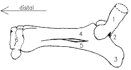 Beef
shankbones showing: 1, distal end of humerus; 2, olecranon fossa; 3,
olecranon process;, 4,radius; 5, ulna; and 6, carpal bones.
The radius is joined to the ulna and is the shorter and more
anterior bone of the pair.
- Beef and lamb carcasses have a set of six compact carpal bones
remaining on the carcass after slaughter. Before slaughter,
the forefeet of cattle and sheep have a large cannon bone located
distally to the carpal bones.
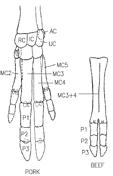 A large plan of the pork foot
compared to a small plan of beef, showing:
AC, accessory carpal;
C, carpal;
IC, intermediate carpal;
MC, metacarpal;
P, phalanges;
RC, radial carpal;
and UC, ulnar carpal.
Beef cannon bones are removed with the feet at slaughter since
there is virtually no meat on them. Cannon bones sometimes are left on
lamb carcasses in the abattoir to prevent the meat contracting
proximally up the limb. Each forelimb cannon bone in ruminants is
derived by the enlargement and fusion of the third and fourth
metacarpal bones.
- In the human hand, the metacarpal bones lie in the flat part
of the hand, between the wrist and the knuckles. If the human hand
is placed flat on a desk and is slowly lifted from the wrist, the
thumb (digit 1) is the first digit to leave the desk, followed by
the index finger and the little finger (digits 2 and 5,
respectively). The third and fourth fingers remain on the desk.
This demonstrates how the feet of meat animals may have evolved
from an original basic plan with five digits.
- In pigs, digits 3 and 4 on each foot bear most of the body
weight and are larger than the lightly loaded digits 2 and 5. The first
digit is absent in pigs. The evolutionary trend towards
lifting of the foot and reduction of digits is even more extensive
in cattle and sheep.
- Cattle and sheep have cursorial limbs, long limbs adapted for
running. Digits 2 and 5 are reduced to dew claws behind the
fetlock. Weight-bearing digits 3 and 4 are enlarged, and their
metacarpals are fused to form a long cannon bone.
- The small bones in the toes of both fore and hind feet are
called phalanges.
- The feet remain on pork carcasses when they are shipped from
the abattoir in shipper's style (unsplit carcasses with head plus
perirenal or leaf fat) or in packer's style (split sides with leaf
fat and head but not jowls removed). However, in a Wiltshire side
for bacon production, the feet are removed, together with the head,
pelvis, vertebral column and psoas muscles.
 Atlas (on left) and axis of
sheep.
 Atlas (on left) and axis of
sheep.

 Plan of neck in beef, showing:1,
ligamentum nuch; 2, atlas; and 3, axis.
The ligamentum nuchae is pale yellow
  with a thick cord-like or
funicular part and a flat sheet-like or lamellar part. Once the
head is removed at slaughter, the elasticity of the ligamentum
nuchae causes the neck of the carcass to curve dorsally.
 Sheep
Sheep Rib showing : 1, head; 2, neck;
3, tubercle; and 4, costal cartilage.Each rib has a tubercle
that articulates
with the transverse process of the more posterior of its two
vertebrae. Ventrally, the anterior ribs articulate with the sternum
and are termed sternal ribs. The more posterior ribs are
called asternal ribs and they only connect to the sternum
indirectly via costal cartilages. The most posterior ribs have only
small costal cartilages that do not reach all the way to the
sternum. Some of the costal cartilages are very hard and may appear
more like bones than typical cartilage.
 This
is the shape of the blade bone in beef.
The scapula is not fused to the vertebral column (like the
pelvis in the
hindlimb), and this allows muscles that hold the scapula to the
ribcage to function as shock absorbers during locomotion.
 Proximally, the humerus has a relatively flat knob or head to fit
into the glenoid cavity of the scapula. Two well defined
condyles on the distal end of the humerus contribute to the hinge
joint at the elbow.
 Beef
shankbones showing: 1, distal end of humerus; 2, olecranon fossa; 3,
olecranon process;, 4,radius; 5, ulna; and 6, carpal bones.
 A large plan of the pork foot
compared to a small plan of beef, showing: