Radial Growth
The radial dimension of the muscle fiber has received much attention
over the years, and the belief that diameters are positively correlated
with meat yield and negatively correlated with meat quality still persists.
But seldom are the intricacies of muscle structure taken into account.
The ends of muscle fibers are rounded if they insert into tendons but tapered
if they insert intrafascicularly beneath the endomysium
of another fiber. As far as we know at present, only rarely are the fibers
of carcass muscles branched at their ends, although branching may occur
in the cutaneous and tongue muscles. Branched fibers also may occur in
diseased and overloaded muscles.
Muscle volume
Muscle volume is almost, although not exactly, constant during contraction.
Thus, as fibers shorten in length, their radial dimensions increase. But
even when fibers have set in rigor mortis, their radii are not stable.
Rigor development is accompanied by a decrease in fiber diameter
as myofilaments are drawn closely together by the active heads of myosin
molecules. Fibers also may lose water as their pH declines towards
the isoelectric point of myofibrillar proteins. If fibers are removed
from a muscle prior to the onset of rigor mortis, they may contract in
a manner that is difficult to control. Even if a muscle sample is clamped
at a predetermined length, there is no guarantee that some parts of the
sample will not contract strongly enough to stretch other parts. The measurement
of radial fiber dimensions must be coupled with some method to reduce all
data to a common muscle length related to sarcomere length. Histological
fixation introduces an extra set of experimental variables. Fixation is
necessary to preserve tissue structure, but some fixatives cause tissue
shrinkage while others cause expansion.
Increase in myofibrils
The radial growth of muscle fibers is associated with an increase in the
apparent number of myofibrils seen in cross section. The apparent number
of myofibrils is a useful measure of radial growth, even though it is rather
an artificial parameter. Ribosomes are present within the myofilament lattice
where presumably they are able to synthesize new myofibrillar proteins.
As new myofilaments are added and the myofibril increases in diameter,
myofibrils may split longitudinally, but seldom are the splits continuous
along the complete length of a muscle fiber. Thus, what looks like one
myofibril in a plane above the split may appear as two myofibrils in a
lower plane. Spitting may occur first at Z lines, where peripheral thin
filaments tend to pull at an angle during contractio.Splitting starts in
the center of an I band and then spreads to the A band and the periphery
of the myofibril, although this is only characteristic of chronic stretching.
Increase in sarcoplasm
As a muscle fiber accumulates contractile proteins during growth, it also
increases its sarcoplasmic volume sarcoplasm and number of mitochondria.
Mitochondria are very abundant in the sarcoplasm of fibers from young animals
but the proliferation of mitochondria may lag behind the increase in sarcoplasmic
volume which occurs with fiber growth. This is most evident in white fibers.
Mitochondria appear to proliferate by fission after each mitochondrion
has been internally subdivided by the formation of a septum.
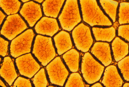
Difficulty of measuring fiber radius
The cross sectional areas of muscle fibers are increased if the fibers
are cut obliquely rather than in exact cross section. Oblique sections
may be caused by poor alignment of the tissue on the microtome chuck but,
even in correctly aligned tissue, they may occur if some fibers of the
sample are contracted more than others. Fibers with a lesser degree of
contraction may be thrown into sinuous folds that give rise to oblique
sections at intervals along the fiber length. If fibers are cylindrical,
this is not a problem since the minimum diameter gives the true diameter.
But if fibers tend are polygonal, minimum diameters may underestimate true
mean diameters.
Giant fibers - are really
big!