4 Embryology of Poultry
4.1 Blastodisc
The main feature is there is very little space available
inside the shell of the egg. Thus, development starts as a flat disc on
top of the yolk.
- Cleavage occurs as the zygote is passing
along the oviduct before the egg is laid.
- The cytoplasm is concentrated
in the blastodisc at the
opposite
pole to the yolk mass.
- The blastodisc is a
pale area
several millimeters across.
- A flat cavity called either the blastocoele
or the
cleft space develops within the depth of the blastodisc to produce
superficial
(epiblast) and deep (hypoblast) layers of cells.
- The
presence of a subgerminal cavity
gives
the central part of the blastodisc a
semitransparent
appearance and this area is called the area
pellucida.
- The
surrounding area
where the edges of the blastodisc are in contact with the yolk is
called the
area opaca.
- All the cells for the future chick embryo are recruited
from the
two layers of cells (epiblast and hypoblast) of the area pellucida.
- The
roles
of cells from various regions of the epiblast and hypoblast in the
developing
embryo have been determined by marking the cells with nontoxic dyes to
produce
a fate map. A simplified fate
map for the epiblast is shown below.
- The
hypoblast produces endoderm,
with a small contribution to
the notochord.

- The epidermis will
become the outer layer of the
skin.
- Neural
tissue will give rise to the brain, spinal cord and nerves.
- The notochord will start
the
development of the vertebral column, and
eventually become cartilaginous intervertebral discs once the
bones of the vertebral column develop.
- Somite will form blocks
of
mesoderm alongside the vertebral column, from which will later develop
vertebrae, muscles and deep tissues of the dermis.
- The lateral plate
mesoderm will develop into the walls of the body cavity.
4.2 Gastrulation

- Gastrulation is
relatively simple in
oligolecithal eggs, but in the flat blastodisc of poultry eggs
gastrulation is
more complex and the cellular intrusion characteristic of gastrulation
occurs
along a slit called the primitive
groove.
- The cells of the primitive
streak sink
downwards and migrate sideways. The primitive streak
develops after
the egg is laid and by about 16 hours of incubation is the most
conspicuous
feature of the developing embryo. The primitive streak indicates the
location
of the future vertebral axis.
- The mesoderm develops
from a middle layer
of
cells accumulating and spreading sideways below the primitive groove.
- The embryo is lifted off the yolk by the
development of infoldings below the future head and tail regions of the
embryo.
These infoldings unite along the sides of the embryo. Thus,
connection to
the yolk is progressively restricted.
- The amnion
grows up and over, all around the embryo, and the amniotic
folds then fuse to form a tent‑like roof over the embryo.
- The notochord
establishes the
future
axis
of the vertebral column and is formed from some of the mesodermal cells
in a
middle layer between the ectoderm and the endoderm along the primitive
streak. Formation of the
notochord starts anteriorly and then posterior
epiblast cells
are recruited as the notochord grows posteriorly.
- Not all of the
mesoderm
within the primitive streak is used for notochord formation and along
each side
of the notochord there remains a long strip of mesoderm developing
into somitic and intermediate mesoderm.
- Laterally, the remaining mesoderm
splits into
outer somatic mesoderm and
inner splanchnic mesoderm. By
this time, the
nerve
cord has developed from a sunken roll of ectoderm sunken located above
the
notochord.
4.3 Induction
Much of our knowledge of animal development comes from the
study of the readily
accessible
embryos of lower vertebrates. It has been known for many
years
that the zygote of the frog exhibits a grey crescent caused by exposure
of
the lower layers of cytoplasm when the surface layers are dragged
inwards by
penetration of a spermatozoon. The grey crescent later becomes the
dorsal lip
of the blastopore - the channel that opens into the central
cavity or
archenteron of the gastrula.
The dorsal lip of the blastopore
eventually forms
part of the inside layer of the gastrula (the archenteron roof) and
later becomes the notochord. The prospective notochord then induces the
formation of
the nerve cord. Many
different parts of the body develop by induction. Embryonic
induction may be caused by ribonucleoproteins.
4.4 Competence
The
tissue responding to induction (the ectoderm in the case of neural tube
formation) must exhibit competence. Inappropriate tissues lacking competence do
not normally exhibit a response to inducers. Thus, the prospective
notochord
cannot induce the formation of a nerve cord in an inappropriate layer
of cells
such as mesoderm. Only ectodermal cells exhibit competence for the
formation of
the nerve cord.
- Competence also may be stage-specific, so only the
appropriate tissue at an appropriate stage of development will respond.
For
example, only the ectoderm of the gastrula may be induced to
form a
nerve cord.
- Regional specificity also may be apparent, as when the dorsal lip
develops in
an anterior to posterior sequence with the older tissue moving downward
and
forward. Thus, the early dorsal lip induces the formation of the
anterior part
of the brain while the late dorsal lip induces the formation of the
posterior
part of the brain.
- As far as is known, inducers work by
activating
genes already present in the target tissue, rather than by
providing
any new or extra genetic information.
4.5 Chick at 24 hours
Although the chick is developing from anterior to posterior
(from top to bottom of the micrograph below), you can see the primitive
streak is posterior. Hensen's node is the edge where the
cells are rolling downwards and anteriorly. The area opaca is the general mottled
area all around the developing embryo where it is resting on the yolk,
whereas the area pellucida is
where the embryo is lifted off the yolk above the subgerminal cavity. Neural folds are visible where the
ectoderm is starting to form the neural tube (leaving an anterior open
end at the neuropore). On each
side of the neural tube are blocks of somitic
mesoderm. The notochord
is faintly visible (it is ventral to the developing neural tube).

The neural tube develops as a roll of ectoderm sinking
downwards into the embryo.
4.6 Chick at 33 hours
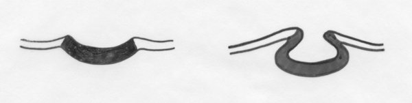
The three main parts of the brain are starting to swell up (prosencephalon, mesencephalon and rhombencephalon). The optic vesicle will become the eye.
The heart seems strangely anterior, but this is because it is still
developing from a tubular structure. The vitelline vein is starting to invade
the yolk to bring in nutrients for the expanding embryo. The process
whereby somites develop from clumped parts of the paraxial mesoderm is visible.

4.7 Chick at 56 hours
Torsion has occurred
so the chick's head is resting sideways on the yolk instead of
face-down into the yolk. The two main parts of the
rhombencephalon (metencephalon
and myelencephalon) have
developed.

Further information
Libraries contain many excellent embryology texts - so grab
the first one you find.
S.F. Gilbert & A.M. Raunio (1997) Embryology.
Constructing the Organism. Sinauer Associates, Sunderland, MA.
This is an amazing book covering the development of many phyla
of animals. The birds and mammals each have their own chapter - thus,
giving the main features in the fewest words.
Trivia
The water colour diagrams originate from my own student notes
which were made largely from ,
B.I. Balinsky. (1965). An Introduction to Embryology. W.B.
Saunders, Philadelphia.
The micrographs were made from whole mounts of chick embryos
stained with carmine (which is why they are red).