16 Fibrous connective tissues
16.1 Introduction
- Fibrous connective tissue are very important in meat - because
they hold the myofibres together.
- Some muscles have extremely strong connective tissue to prevent
the myofibres being damaged as the muscle contracts.
- Muscle with an angular arrangement of myofibres gain leverage
when they contract. The length of the contraction is reduced but
the strength is increased. The myofibres must be anchored in strong
connective tissue.
- The strongest connective tissues are found in the distal muscles of the limbs and in
the neck.
- Muscles with a high connective tissue content must be cooked with
moist heat (stewed not
barbecued or roasted).
- Connective tissues tend to be stronger
in large animals (eg., beef) than in small animals (eg., lamb).
- Connective tissues tend to be stronger
in old animals (eg., mature beef) than in young animals (eg.,
veal).
- This explains why stewing beef (neck and distal limb muscles) is
less expensive than steak or roasts.
- This also explains why beef
carcasses are graded by animal age. Grade A beef is from
youthful animals (up to about 18 months of age).
Here is an image a fibrous connective tissue.

It has three essential features: (1) cells (stained with
methylene blue), (2) fibres (stained pink with eosin), and (3) matrix
(in the spaces between the cells and the fibres).
 The fibrous connective
tissues in meat form a continuous mesh, as shown in the image to the
left,
from the microscopic strands of endomysium
around individual myofibres,
to the thick layers of perimysium
around bundles of myofibres
(fasciculi),
all being gathered and connected to the very thick epimysium on the
surfaces of individual muscles.
The fibrous connective
tissues in meat form a continuous mesh, as shown in the image to the
left,
from the microscopic strands of endomysium
around individual myofibres,
to the thick layers of perimysium
around bundles of myofibres
(fasciculi),
all being gathered and connected to the very thick epimysium on the
surfaces of individual muscles.
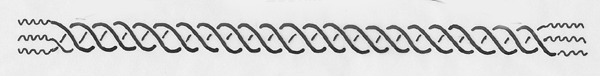
The image below shows a thick layer of perimysium.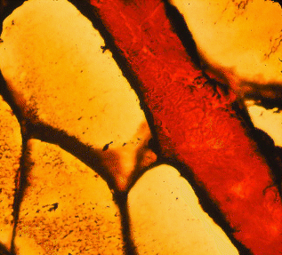
The endomysium, perimysium and epimysium contain two types of
protein fibres, collagen and elastin, which now we will consider
in detail.
16.2 Collagen fibers
- Tropocollagen is an
elongated protein forming extremely strong but very small collagen fibrils
(best seen with an electron
microscope).
- Numerous collagen fibrils
are bound together to form collagen fibres
visible with a light
microscope.
- When collagen fibers form sheets or cables we
can see them in meat without a microscope, and we may detect them
as
gristle if they do not gelatinize when meat is cooked.
- Note: collagen fibres are
extracellular (outside the cells forming them) whereas myofibres are
cells themselves.
- Tropocollagen is the most
abundant protein in the animal body.
- Large amounts of tropocollagen are found in animal skin. In pig
skin, for
example, collagen fibres are tightly woven from two directions to form
a tight meshwork
- Collagen is a raw material for major industries
in leather, glue, cosmetics, food processing (gelatin), etc.
- Under a light microscope, collagen fibres in the connective
tissue framework
of meat range in diameter from 1 to
12 micrometres (0.001 millimetre =
1 micrometre).
- Collagen fibres do not often
branch and, when branches are found, they
usually diverge at an acute angle.
- Collagen fibres from fresh meat are
white, but usually they are stained in histological sections for
examination
under a microscope. The most common stain for light microscopy is
eosin,
which stains collagen fibres pink.
- Unstained collagen fibres may be seen
by polarized light since they are birefringent (with two refractive
indices like A bands). By rotating the plane of polarized
light, collagen fibres appear bright against an otherwise dark
background
(when two Polaroid lenses are perpendicular they block most of the
light,
but collagen fibres can rotate the light so they appear bright).
- The
birefringence of collagen fibres in meat is lost during cooking at the
point gelatinization occurs.
- Collagen fibres have a wavy or crimped appearance
which disappears when they are placed under tension.
- Collagen fibres fluoresce with a blue-white light when
excited
with UV light enabling the amount of connective tissue on a cut meat
surface
to be measured very rapidly. Peak excitation is around 370 nm so that
the prominent 365 nm peak emission of a mercury arc lamp may be used.
Some
indication of collagen fibre diameter may be obtained by
spectrofluorometry
(measuring the wavelengths of fluorescence) because the fluorescence is
quenched (fades) fairly rapidly. Thus, large collagen fibres retain a
central
core with a pre-quenching emission spectrum for longer than small
fibres.
Fat only fluoresces weakly, to about the same extent as areas of muscle
with a low connective tissue content. Collagen fluorescence
increases with animal age.
- Electron microscopy shows collagen fibrils with diameters ranging from 20 to 100 nm (0.001
micrometre = 1 nanometre). Collagen fibrils typically have diameters
which
are multiples of 8 nm showing the manner in which they grow radially.
- Collagen microfibrils
(even smaller structures that make up fibrils) may
appear to have a tubular structure with an electron-lucent lumen
(appearing
empty under the electron microscope).
- Collagen fibrils
are formed from long tropocollagen
molecules which are staggered in arrangement
but tightly bound laterally by covalent chemical bonds.
- For electron microscopy,
when negatively stained with heavy
metals, the stain spreads into the spaces between
the ends of molecules, and collagen fibrils appear to be transversely striated.
- The periodicity of these striations is 67 nm but often shrinks to
64 nm
as samples are processed for examination
- Collagen fibres are formed by cells called fibroblasts.
- Although collagen fibres are
located outside the cell, the initial stages of collagen fibril
assembly may be within the cell, with fibril morphology being
regulated
by a special site on the fibroblast membrane.
Tropocollagen is a high molecular weight protein (300,000
Daltons) formed
from three polypeptide strands
twisted into a triple helix. Each strand
is a left-handed helix twisted on itself, but the three strands are
twisted
into a larger right-handed triple helix. The triple helix is
responsible
for the stability of the molecule and for the property of self-assembly
of molecules into microfibrils. The flexible parts of each strand
projecting
beyond the triple helix (telopeptides)
are responsible for the bonding
between adjacent molecules. In other words, the cross links binding
tropocollagen molecules together laterally are made between the helical
shaft of one molecule and the non-helical extension of an adjacent
molecule.
In the polypeptide strands, the small amino acid glycine
occurs
at every third position,
and proline and hydroxyproline account for 23%
of the total residues. The regular distribution of glycine is required
for the packing of tropocollagen molecules and has been claimed as
evidence all animals are derived by evolution from a single ancestral
stock,
since the chance development of this unique regularity in unrelated
animals
is thought unlikely. Hydroxyproline is quite rare in other proteins of
the body, and an assay for this imino acid (an imino acid is
chemically
similar, but not the same as an amino acid) provides a measure of the
collagen
or connective tissue content in a meat sample. Tropocollagen also
contains
a fairly high proportion of glutamic acid and alanine as well as some
hydroxylysine.
16.4 Biochemical types of collagen
The various types of collagen of interest in understanding the
structure
of meat are as follows.
- Type I collagen
forms striated fibres between 80 and 160 nm in diameter
in blood vessel walls, tendon, bone, skin and meat. It may be
synthesized
by fibroblasts, smooth muscle cells (around blood vessels) and
osteoblasts
(bone-forming cells).
- Type II collagen
fibres are less than 80 nm in diameter and occur in hyaline
cartilage (for example, in the soft blade of the scapula) and in
intervertebral discs. It is synthesized by chondrocytes
(cartilage-forming cells).
- Type III collagen
forms reticular fibres in
tissues with some degree of
elasticity, such as spleen, aorta and muscle. It is synthesized by
fibroblasts
and smooth muscle cells, contributes substantially to the endomysial
connective
tissues around individual myofibres, provides a small fraction of the
collagen found in skin and occurs in the large collagen fibres
dominated
by Type I collagen. It may have some function in regulating collagen
fiber
growth. Unlike Type I collagen fibres, reticular Type III fibres are
highly branched.
- Type IV collagen
occurs in the basement membranes around many types of
cells and may be produced by the cells themselves, rather than by
fibroblasts.
Although basement membranes were once regarded as amorphous (like
glue),
many of them now are thought to be composed of a network of irregular
cords.
The cords contain an axial filament of Type IV collagen, ribbons of heparin sulphate, proteoglycan, and fluffy material (laminin, entactin and fibronectin).
Type IV collagen occurs in the endomysium around individual muscle
fibers.
Instead of being arranged in a staggered array, the molecules are
linked
at their ends to form a loose diagonal lattice.
Tendons often extend into the belly of a muscle or along its
surface before
they merge with its connective tissue framework, and Types I and III
collagen
both may be extracted from meat. Even within tendons, there may be some
Type III collagen forming the endotendineum
or fine sheath around bundles
of collagen fibrils. In fibres composed of collagen Types I and II,
fibrils
have a straight arrangement whereas, in fibres of Type III collagen,
the
fibrils have a helicoidal arrangement.
Small diameter Type III
collagen fibres are called reticular fibers
since, when stained with silver for light
microscopy, they often appear
as a network or reticulum of fine fibres. The larger diameter collagen
fibres formed from Type I collagen are not blackened by silver.
Collagen fibres shrink when they are placed in hot water, and
ultimately
they may be converted to gelatin. Around 65ºC, the triple helix
is disrupted and the alpha chains fall into a random arrangement. The
importance
of this change? It tenderizes meat with a high connective tissue
content.
Tropocollagen molecules from older
animals are more resistant
to heat disruption than those from younger animals.
16.4
Collagen biosynthesis
The synthesis of the different polypeptide strands combining
to
make different types of tropocollagen is genetically regulated by the
production
of messenger RNA. The synthesis of polypeptide strands occurs on
membrane-bounded
polysomes, but the hydroxylation
of lysine and proline occurs after the
strands are assembled. Ascorbic acid
is required for the hydroxylation
of lysine and proline. Polypeptide strands enter the cisternae
of the endoplasmic
reticulum (a membranous assembly labyrinth within the cell), the
terminal
extensions of the strands are aligned, and then the strands spiral
around
each other. Procollagen or
immature tropocollagen has long terminal extensions
protruding from each end of the newly formed triple helix. Procollagen
moves to the golgi apparatus
and is packaged into vesicles moved
to the cell surface, probably by microtubules. Except for some Type III
procollagen molecules, the long terminal extensions are then
enzymatically
reduced in length.

Outside the cell, tropocollagen molecules become aligned in parallel formations,
and then they link up laterally to form fibrils. It is likely that
tropocollagen
monomers are partially assembled together in groups before they are
added
to an existing collagen fibril. Firstly, vacuoles containing
procollagen
fuse to form a fibril-containing compartment. Then the cytoplasmic
extensions
withdraw from between several fibril-forming compartments to create a
bundle-forming
compartment.
16.5 Crosslinking of tropocollagen molecules
- Within an individual tropocollagen molecule, the three
polypeptide strands are
linked together by stable intramolecular bonds
originating in the non-helical
ends of the molecule.
- The great strength of collagen
fibres, however, originates mainly
from the stable intermolecular
covalent bonds between adjacent tropocollagen
molecules.
- Stable disulphide bonds between cystine molecules in the triple
helix also
occur.
- During the growth and
development of meat animals, covalent
cross
links increase in number, and collagen fibres
become progressively stronger.
- Meat from older animals,
therefore, tends to be tougher than meat from
the same region of carcasses from younger animals.
- This relationship is
complicated in young animals by the rapid synthesis of large amounts of
new tropocollagen. New tropocollagen has fewer cross links so, if there
is a
high proportion of new tropocollagen, the mean degree of cross linking
may be
low, even though all existing molecules are developing new cross links.
- As the formation of new tropocollagen slows down, the mean degree
of cross linking
increases.
- Another complication is many of the intermolecular cross
links in young animals are reducible
(the collagen is strong but is fairly
soluble).
- In older animals, reducible
cross links are converted
to non-reducible cross links (the collagen is strong but is far
less soluble
and more resistant to moist heat).
- Pyridinoline is a non-reducible cross-link causing
increased heat stability of connective tissues from older
animals. It is formed without enzymes by glycosylation (a reaction
between lysine and reducing sugars).
- Differences in the degree of cross linking may occur between
different
muscles of the same carcass, and between the same muscle in different
species.
- Nutritional factors such as high-carbohydrate diet, fructose
instead
of glucose in the diet, low protein, and pre-slaughter feed restriction
may reduce the proportion of stable cross links.
- The rate
of collagen turnover in skeletal muscle may be about 10% per day and
the
turnover time for collagen may be inversely proportional to collagen
fibril
diameter.
16.6 Elastic fibres & elastin
Individual collagen fibres only lengthen by about 5% when
stretched and
little elasticity is possible where collagen is formed into cable-like
tendons. However, much of the collagen present in meat forms a
meshwork and stretching of the whole meshwork is possible because its
configuration changes. Fibres with truly elastic properties, however,
are
necessary in structures such as the
ligamentum nuchae of the neck and in the
abdominal wall. And all arteries,
from the aorta down to the finest microscopic
arterioles, rely on elastic fibres
to accommodate the surge of blood from
contraction of the heart. Elastic fibres may be stretched to several
times
their original length but rapidly resume their original length once
released.
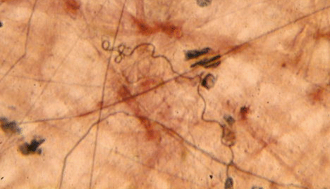
Elastic fibres are composed of the protein, elastin. Elastin is found in all
vertebrates except primitive jawless fish, and
in evolution it appeared first in cartilaginous fish. The elastic
fibers
in the image below (from loose connective tissue around the intestine)
are the thin black ones. The much thicker, red-brown fibres are
collagen.
Elastin resists severe chemical conditions, such as the extremes of
alkalinity,
acidity and heat destroying collagen.
- Fortunately, there are
relatively few elastic fibres in muscle, otherwise
cooking would do little to reduce meat toughness.
- The elastic fibres in
muscles used frequently for locomotion are larger and more
numerous
than those of less frequently used muscles.
- Elastic fibres in the epimysium
and perimysium of beef muscles range from 1 to 10 microns in diameter.
- Elastic fibres usually are pale yellow.
- When elastic fibres are stretched,
they may become visible in polarized light without staining.
- Elastic fibres in the connective
tissue framework of meat are usually branched.
- Electron microscopy reveals elastic fibres are composed of
bundles
of small fibrils approximately 11 nm in diameter embedded in an
amorphous
material.
- Although elastin resembles tropocollagen in having a large amount
of
glycine, it is distinguished by the presence of two unusual amino
acids, desmosine and isodesmosine.
- Like collagen, elastin contains hydroxyproline,
although it may not have the same function of stabilizing the molecule.
- Tropoelastin, the soluble
precursor molecule of elastin (molecular weight
70,000 to 75,000 Daltons), is secreted by fibroblasts after it has been
synthesized
by ribosomes of the rough endoplasmic reticulum and processed by the
Golgi
apparatus.
- In the presence of copper,
lysyl oxidase links together four
lysine molecules to form a desmosine molecule.
- Isodesmosine is the isomer
of desmosine.
- The aorta may be fatally weakened by a lack of mature elastin
in animals deprived of dietary copper.
- Elastin in the arterial system is
produced by smooth muscle cells instead of fibroblasts.
16.7 Why meat must be a natural food for us
During the digestion of meat in
the human gut,
elastic fibres are broken down by elastase,
an enzyme from the pancreas.
We would not have this enzyme if our evolutionary ancestors had not
been at least
partly carnivorous. In other words, I have never read of the occurrence
of elastin in any human food except meat. So if we have evolved a
highly
specific enzyme, elastase, to deal with elastin in our food, this can
only
mean that we are the descendants of meat eaters.
Further information
Structure and Development of Meat
Animals and Poultry. Pages 82-95.