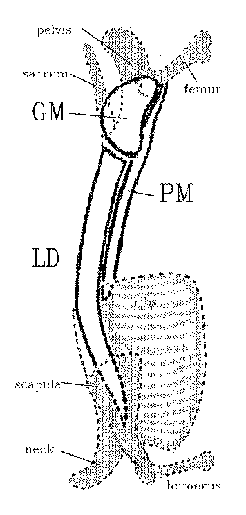
At the
end of LAB 9 we reached as far back as the loin
muscles. We will repeat these again because of their
importance, and then continue into the hindquarter. Remember LD
is the longissimus dorsi, PM
is the psoas major and GM is
the gluteus medius.

The loin muscles give rise to tender
meat
with a desireable taste, and they command a high price when presented
for sale
as steaks or chops. The longissimus dorsi extends posteriorly from the
rib
region, it runs through the loin, and most of the muscle terminates on
the
anterior face of the ilium. Thus, the longissimus dorsi is seen in cuts
of meat
taken through the ribs and loin, but not in cuts of meat such as
sirloin steaks
that are posterior to the anterior face of the ilium. The longissimus
dorsi is
dorsal to the transverse processes of the lumbar vertebrae, and it is
dorsal to
the ribs in the thoracic region. For most of the length of the ribcage,
there
are no major muscles immediately ventral to the heads of the ribs.
Thus, in a
rib steak, there is only one main eye of meat, and that is the
longissimus
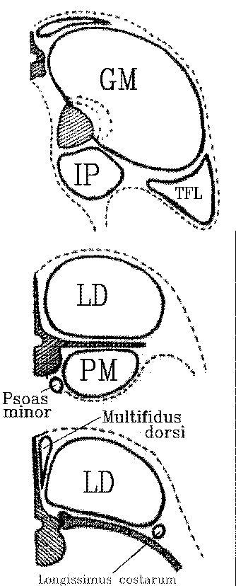
dorsi dorsal to the ribs. However, in the loin, there are muscles both
above
and below the level of the transverse processes of the lumbar
vertebrae. The
dorsal muscle above the transverse processes is the longissimus dorsi.
The
ventral muscle below the transverse processes is the psoas major or
filet
muscle. The psoas major originates ventrally to the last couple of ribs
in the
ribcage. The cross sectional area of the psoas major increases towards
the
sirloin. Medial to the psoas major, almost under the centra of the
vertebrae,
is a smaller psoas muscle called the psoas minor. The letter P in the
word
psoas is silent when the word is spoken.

Immediately lateral to the dorsal spines
of
the vertebrae (medial to the longissimus dorsi) are some small
multifidus dorsi
muscles. The longissimus costarum is a relatively small rope‑like
muscle,
dorsal to the ribs. It appears as a small eye of meat at the separation
between
the forequarter and the hindquarter. The multifidus dorsi and the
longissimus
costarum have little commercial significance, since they are such small
muscles, but they may create a problem in the measurement of rib‑eye
areas
since they might be accidently included with the longissimus dorsi.
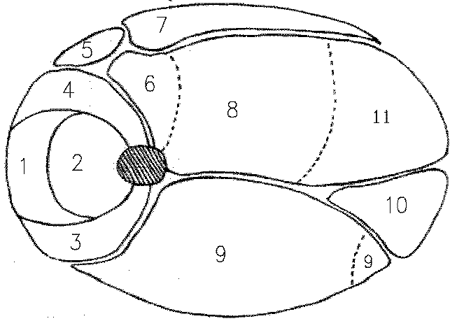
The muscles of the hindlimb are
relatively
large and produce a large volume of moderately tender meat. The
gracilis is a
thin sheet of muscle spread over the medial face of the hindlimb. The
gracilis,
together with the small sartorius muscle anterior to the gracilis, may
be used
to orient hindlimb cuts of meat such as a beef round steak or a slice
of ham.
Orientation is necessary for the identification of the remaining
muscles of the
hindlimb which are grouped around the femur. The quadriceps femoris
muscles
form a group of four large muscles that pull on the patella when the
leg is
extended. The vastus medialis is medial, the vastus lateralis is
lateral, the
vastus intermedius covers the anterior face of the femur, and the
rectus
femoris covers the vastus intermedius.

The biceps femoris is a single large
muscle
on the lateral face of the hindlimb. In cross section, it often appears
to be
divided into two parts because it has a very deep cleft along part of
its
length. The biceps femoris appears as a single muscle in cuts of meat
which
miss the cleft, but sections through the cleft make the muscle appear
double.
To add to the possibility of confusion, the small segment of muscle cut
off by
the cleft is often paler than the main part of the muscle.

The semitendinosus and semimembranosus
are
two large muscles located on the posterior face of the hindlimb. The
semimembranosus is medial to the semitendinosus. The adductor and
pectineus are
located in the medial part of the hindlimb, near to the femur. The
pectineus is
anterior to the adductor. In lean carcasses, it may be difficult to
separate
the adductor from the semimembranosus, and these two muscles may appear
as a
single muscle. Given the possibility that the biceps femoris muscle on
the
other side of the limb may look like two muscles, caution
is needed in the identification of these muscles for the
first time. The gastrocnemius is a large muscle located deep in the
hindlimb
and is covered by distal extensions of
some of the proximal muscles of the limb. The gastrocnemius pulls on
the
Achilles tendon at the hock, and is the equivalent of the calf muscle
in the
human leg. A major anatomical difference between the human leg and the
hind
limbs of meat animals is that the human gastrocnemius is not covered by
the
posterior thigh muscles.
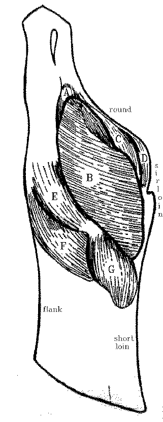
Between the hindlimb and the loin, and
located laterally to the pelvis, are several large muscles that form
the rump
and sirloin of the carcass. The positioning and naming of the first of
these
muscles is rather misleading. Despite its name, the gluteus medius is
located
laterally in meat animals. It covers the lateral face of the ilium and
appears
as the large muscle area in sirloin steaks and chops. From lateral to
medial,
there are three layers of muscles, gluteus medius, gluteus accessorius
and gluteus
profundus. The gluteus medius is the largest of the three.

The psoas major continues posteriorly
from
the loin into the sirloin. It is joined by another muscle, the iliacus,
and the
two together may be given a compound name, the iliopsoas. Little, if
any, of
the longissimus dorsi appears in the sirloin since most of the muscle
terminates on the anterior face of the ilium.