 Notice
the cerebellum
at the bottom right corner. It is darker in color and like a small
version of the cerebellum.
Notice
the cerebellum
at the bottom right corner. It is darker in color and like a small
version of the cerebellum.Cerebrum - L & R cerebral hemispheres
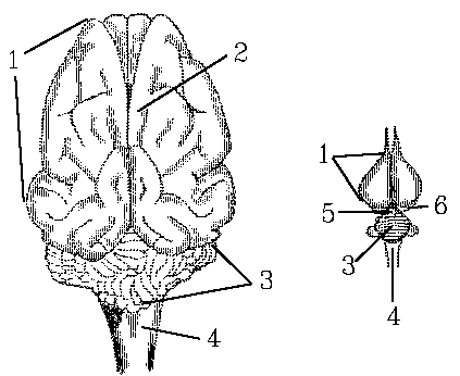
Cerebellum
Gyri & sulci
Medulla oblongata - control of heart & respiration
Epiglottis
Turbinate bones, rolled up in nasal cavity
Ventricle - brain cavity, connects with cerebrospinal fluid cavity
Thalamus - hypothalamus - pituitary
Meninges - cover brain
The nervous system is a single integrated system composed of distinct regions.

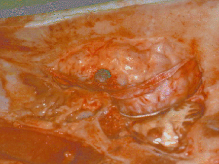
This image shows a side view of a pig brain, with the skull split
longitudinally. The dark area was caused by the placement of an
electrode in an experiment undertaken to find ways to improve animal
slaughter methods. Notice
the cerebellum
at the bottom right corner. It is darker in color and like a small
version of the cerebellum.
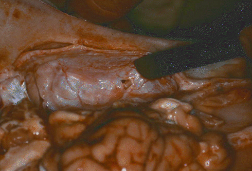
The surface of the brain is covered by a delicate membrane (pia mater) that carries a network of small blood vessels supplying the brain. The pia mater is covered by another thin membrane called the arachnoid membrane. On top of this membrane is a layer of tough tissue, the dura mater, that adheres to the inner surface of the skull. In the image above, you can see that these membranes are being peeled off the surface of the brain towards the bottom right corner. In the image below, the dark ruler is showing the dura mater still adhering to the inside of the cranial cavity.

The surface of the brain in meat animals is increased in area by folds and grooves called, respectively, (gyri) and (sulci).
The cerebrum is composed of left and right cerebral hemispheres separated by a deep fissure. If the brain is exposed by cutting through the skull with a band saw, the outer layers of the brain appear gray in color while the inner parts appear white. The gray areas are dominated by nerve cell bodies while the white areas are dominated by axons. Axons are cable-like extensions of the nerve cell body or (perikaryon), and they are electrically insulated by a sheath of myelin.
The gyri and sulci on the surface of the brain allow large numbers of nerve cell bodies to connect with the bundles of axons which carry information within the central nervous system.
The function of the cerebrum is to regulate higher forms of nervous activity such as recognition, learning, communication and behavior.
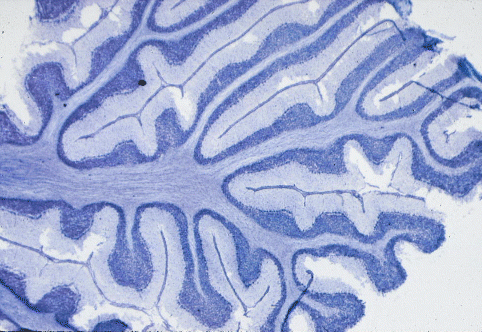
The cerebellum is posterior to the cerebrum, and is formed from a middle lobe called the vermis and two lateral hemispheres. The cerebellum coordinates muscle movements during locomotion and in the maintenance of posture. Its internal architecture shown below matches up to the gyri on its surface.

At higher magnification, some of the largest neurons are visible.

The thalamus is the region of the brain located ventrally to the cerebrum. It links the cerebrum to the rest of the central nervous system. The hypothalamus is located ventrally to the thalamus and connects the major regulatory gland of the endocrine system, the pituitary, to the brain.
The most posterior region of the brain, where it tapers down to the diameter of the spinal cord, is called the medulla oblongata. This region controls the heart rate via elements of the autonomic nervous system described earlier.
Cavities or ventricles filled with cerebrospinal fluid run through the brain and extend down the spinal cord as a small canal.

The spinal cord is an extension of the brain. It emerges from the skull through the foramen magnum, and extends posteriorly to the first (cattle and sheep) or third (pigs) sacral vertebrae in the sirloin region.
At regular intervals, pairs of dorsal and ventral roots enter and leave the spinal cord. Dorsal and ventral roots unite outside the spinal cord to form the nerves of the peripheral nervous system. The nerves that are visible in meat are composed of very large numbers of microscopic axons.
Sensory neurons with their cell bodies in the dorsal root ganglia at the side of the spinal cord carry incoming sensory information from the skin, muscles and tendons. Sensory axons terminate on a variety of different types of neurons in the spinal cord. These neurons may relay information up the spinal cord to the brain, or to other regions of the spinal cord, or to nearby motor neurons.
Motor neurons have their cell bodies in the ventral horns of the central gray region of the spinal cord. In a simple spinal reflex, motor neurons are activated via the dorsal root by axons from sensory neurons). Motor axons leave the spinal cord in ventral roots and innervate muscle fibers in the carcass muscles. Motor axons do not branch much before they reach their muscles but, once inside a muscle, they branch extensively to innervate a large group of muscle fibers called a motor unit. The synapse at which a terminal branch of an axon connects to a muscle fiber is called a MOTOR END PLATE.
Myelinated axons which ascend and descend the spinal cord are
located outside the central gray areas so that,
The spinal cord is located within the vertebral column. It is separated from the inner bony surfaces of the vertebrae by an epidural space.
ates glands and viscera.